
Page 475 - Adams and Stashak's Lameness in Horses, 7th Edition
P. 475
Lameness of the Distal Limb 441
VetBooks.ir
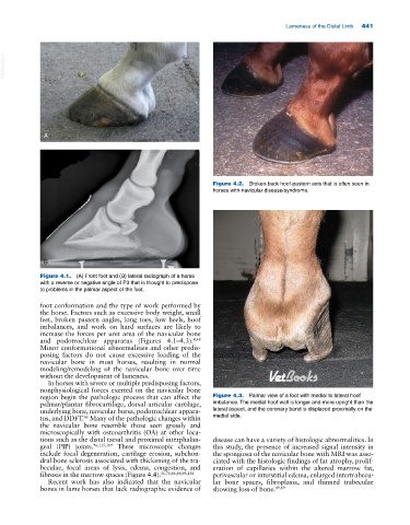
A
Figure 4.2. Broken‐back hoof‐pastern axis that is often seen in
horses with navicular disease/syndrome.
B
Figure 4.1. (A) Front foot and (B) lateral radiograph of a horse
with a reverse or negative angle of P3 that is thought to predispose
to problems in the palmar aspect of the foot.
foot conformation and the type of work performed by
the horse. Factors such as excessive body weight, small
feet, broken pastern angles, long toes, low heels, hoof
imbalances, and work on hard surfaces are likely to
increase the forces per unit area of the navicular bone
and podotrochlear apparatus (Figures 4.1–4.3). 4,48
Minor conformational abnormalities and other predis-
posing factors do not cause excessive loading of the
navicular bone in most horses, resulting in normal
modeling/remodeling of the navicular bone over time
without the development of lameness.
In horses with severe or multiple predisposing factors,
nonphysiological forces exerted on the navicular bone
region begin the pathologic process that can affect the Figure 4.3. Palmar view of a foot with medial to lateral hoof
palmar/plantar fibrocartilage, dorsal articular cartilage, imbalance. The medial hoof wall is longer and more upright than the
underlying bone, navicular bursa, podotrochlear appara- lateral aspect, and the coronary band is displaced proximally on the
tus, and DDFT. Many of the pathologic changes within medial side.
96
the navicular bone resemble those seen grossly and
microscopically with osteoarthritis (OA) at other loca-
tions such as the distal tarsal and proximal interphalan- disease can have a variety of histologic abnormalities. In
geal (PIP) joints. 96,115,119 These microscopic changes this study, the presence of increased signal intensity in
include focal degeneration, cartilage erosion, subchon- the spongiosa of the navicular bone with MRI was asso-
dral bone sclerosis associated with thickening of the tra- ciated with the histologic findings of fat atrophy, prolif-
beculae, focal areas of lysis, edema, congestion, and eration of capillaries within the altered marrow fat,
fibrosis in the marrow spaces (Figure 4.4). 35,70,86,88,96,136 perivascular or interstitial edema, enlarged intertrabecu-
Recent work has also indicated that the navicular lar bone spaces, fibroplasia, and thinned trabeculae
bones in lame horses that lack radiographic evidence of showing loss of bone. 49,69