
Page 477 - Adams and Stashak's Lameness in Horses, 7th Edition
P. 477
Lameness of the Distal Limb 443
Clinical Signs of lameness in a recent study. The remainder of the
100
horses had a unilateral lameness until a PD nerve block
Horses with navicular disease/syndrome usually have
VetBooks.ir a history of progressive, chronic, unilateral, or bilateral the opposite forelimb. 84,100,101 However, the true severity
was performed, at which time the lameness switched to
forelimb lameness, which may have an insidious (most
of a unilateral lameness at a straight trot may be misin-
35,84,115
common) or acute onset.
The history may include
a gradual loss of performance, stiffness, shortening of terpreted because of the concurrent lameness in the
opposite forelimb. In addition, the horse may tend to
the stride, loss of action, unwillingness to turn, and point one forelimb or alternate pointing each fore-
increased lameness when worked on hard surfaces. 35,46 limb. 115,135 Asymmetry in the extensor muscles with
Chronic bilateral forelimb lameness is considered the atrophy of the muscles associated with the lame limb
norm, and navicular syndrome has been described as can often be observed in horses with chronic
heel pain that blocks to a PD nerve block on both fore- lameness. 115,116,135
limbs with or without radiographic abnormalities. 100,101 In a recent study that compared specific sources of
However, unilateral lameness can also occur, especially foot lameness verified on MRI (navicular bone lesions
with lesions that involve the flexor surface of the navic- alone, lesions of the podotrochlear apparatus, lesions of
ular bone and/or the DDFT. Most horses with an acute the DDFT alone, and lesions involving navicular bone
onset of unilateral lameness that blocks to the foot are and soft tissue), there were no significant associations
not considered to have navicular disease/syndrome, and among groups with respect to onset of lameness, foot
lesions in the DDFT or other problems in the foot should conformation, posture, presence of distal interphalan-
be suspected. geal (DIP) joint effusion, hoof tester examination, flex-
Navicular disease/syndrome is considered to be a ion testing, or blocking pattern. Most (73.3%) horses
84
degenerative process due to wear and tear similar to were lamer when lunged on a firm surface than a soft
OA; therefore, middle‐aged to older horses are most surface, and 32.7% of horses were lamer on a circle
commonly affected. Clinical signs usually become than in a straight line. Horses with podotrochlear appa-
apparent in most horses between 7 and 10 years of age, ratus injuries were more likely to be unilaterally lame,
although younger horses can be affected. 35,46,100,115 and horses with injury to the DDFT were more likely to
Horses with developmental/congenital abnormalities exhibit lameness at the walk when turning tight turns. 84
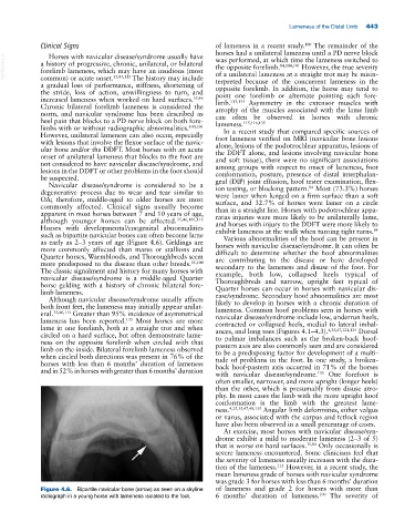
such as bipartite navicular bones can often become lame Various abnormalities of the hoof can be present in
as early as 2–3 years of age (Figure 4.6). Geldings are horses with navicular disease/syndrome. It can often be
more commonly affected than mares or stallions and difficult to determine whether the hoof abnormalities
Quarter horses, Warmbloods, and Thoroughbreds seem are contributing to the disease or have developed
more predisposed to the disease than other breeds. 35,100 secondary to the lameness and disuse of the foot. For
The classic signalment and history for many horses with example, both low, collapsed heels typical of
navicular disease/syndrome is a middle‐aged Quarter Thoroughbreds and narrow, upright feet typical of
horse gelding with a history of chronic bilateral fore- Quarter horses can occur in horses with navicular dis-
limb lameness. ease/syndrome. Secondary hoof abnormalities are most
Although navicular disease/syndrome usually affects likely to develop in horses with a chronic duration of
both front feet, the lameness may initially appear unilat- lameness. Common hoof problems seen in horses with
eral. 35,46,115 Greater than 95% incidence of asymmetrical navicular disease/ syndrome include low, underrun heels,
135
lameness has been reported. Most horses are more contracted or collapsed heels, medial to lateral imbal-
lame in one forelimb, both at a straight trot and when ances, and long toes (Figures 4.1–4.3). 4,35,47,121,135 Dorsal
circled on a hard surface, but often demonstrate lame- to palmar imbalances such as the broken‐back hoof‐
ness on the opposite forelimb when circled with that pastern axis are also commonly seen and are considered
limb on the inside. Bilateral forelimb lameness observed to be a predisposing factor for development of a multi-
when circled both directions was present in 76% of the tude of problems in the foot. In one study, a broken‐
horses with less than 6 months’ duration of lameness back hoof‐pastern axis occurred in 71% of the horses
and in 52% in horses with greater than 6 months’ duration with navicular disease/syndrome. One forefoot is
135
often smaller, narrower, and more upright (longer heels)
than the other, which is presumably from disuse atro-
phy. In most cases the limb with the more upright hoof
conformation is the limb with the greatest lame-
ness. 4,25,35,47,48,135 Angular limb deformities, either valgus
or varus, associated with the carpus and fetlock region
have also been observed in a small percentage of cases.
At exercise, most horses with navicular disease/syn-
drome exhibit a mild to moderate lameness (2–3 of 5)
that is worse on hard surfaces. 35,84 Only occasionally is
severe lameness encountered. Some clinicians feel that
the severity of lameness usually increases with the dura-
115
tion of the lameness. However, in a recent study, the
mean lameness grade of horses with navicular syndrome
was grade 3 for horses with less than 6 months’ duration
Figure 4.6. Bipartite navicular bone (arrow) as seen on a skyline of lameness and grade 2 for horses with more than
100
radiograph in a young horse with lameness isolated to the foot. 6 months’ duration of lameness. The severity of