
Page 481 - Adams and Stashak's Lameness in Horses, 7th Edition
P. 481
Lameness of the Distal Limb 447
in horses with lameness isolated to the foot but with no Scintigraphy
radiographic abnormalities included changes to the Scintigraphy (nuclear imaging) was initially thought
VetBooks.ir DIP joint (69%), and navicular bursitis (56%). Lesions to be able to identify early pathologic changes within
DDFT (73%), effusion and synovial proliferation to the
94
the navicular bone related to its ability to identify early
of the DDFT were most commonly found in the lateral
lobe of the tendon and located just above the navicular alterations in bone metabolism rather than relying on
anatomic changes. However, increased radiopharmaceu-
bone in the majority of cases. These findings were similar tical uptake (IRU) has been documented to occur in only
to results of previous imaging and histopathologic stud- 36% of the limbs of horses with navicular disease/syn-
ies. 10,11,20,39,42,44 Similar to radiography, the accuracy of drome and in a very small percentage of limbs with soft
ultrasound to document pathology within the foot has tissue injuries within the foot. 38,40,67 Scintigraphic assess-
been criticized based on results of more advanced imag- ment of the foot can be helpful to identify the potential
ing such as MRI. Therefore, negative findings with ultra- source of pain causing lameness, but false‐positive
sound do not rule out the presence of abnormalities in results can occur, especially in horses with low‐heel con-
the navicular region and currently is not used routinely formation. 38,40 Additionally, a negative scintigram of the
clinically.
foot does not preclude significant injuries, and scintigra-
phy does not provide a definitive diagnosis. 38,40,96 Results
of a recent study that evaluated the diagnostic accuracy
of scintigraphy in lame and poorly performing sport
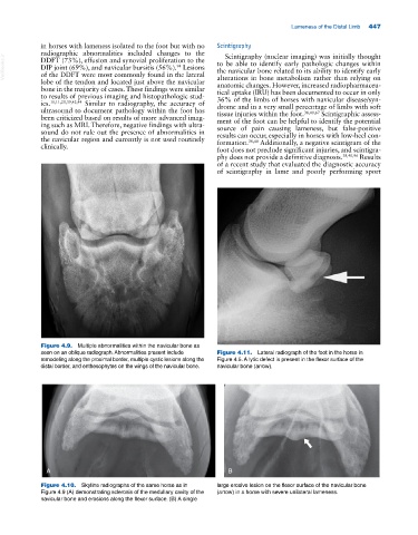
Figure 4.9. Multiple abnormalities within the navicular bone as
seen on an oblique radiograph. Abnormalities present include Figure 4.11. Lateral radiograph of the foot in the horse in
remodeling along the proximal border, multiple cystic lesions along the Figure 4.5. A lytic defect is present in the flexor surface of the
distal border, and enthesophytes on the wings of the navicular bone. navicular bone (arrow).
A B
Figure 4.10. Skyline radiographs of the same horse as in large erosive lesion on the flexor surface of the navicular bone
Figure 4.9 (A) demonstrating sclerosis of the medullary cavity of the (arrow) in a horse with severe unilateral lameness.
navicular bone and erosions along the flexor surface. (B) A single