
Page 480 - Adams and Stashak's Lameness in Horses, 7th Edition
P. 480
446 Chapter 4
mineralization in the DSIL, fracture directly off the more frequently with digital radiography and are thought
distal border of the navicular or fracture of entheso- to be associated with navicular disease (Figure 4.12). 7,8,37
VetBooks.ir 4. Numerous (>7) large and variably shaped distal bor- or dystrophic mineralization within the DSIL. See
phyte formation at the origin of the DSIL.
These fragments may represent a fracture, enthesophyte,
7
der radiolucent zones.
Chapter 4 for further information on radiography of
5. Discrete radiolucent areas in the spongiosa with or the navicular bone.
without detectable communication with the flexor
cortex. Ultrasonography
6. New bone at the sagittal ridge.
7. Increased thickness of the flexor cortex. Ultrasound is an economical and readily available
8. Sclerosis of the spongiosa. diagnostic technique that can be used to help diagnose
9. A bipartite bone. 37 potential soft tissue injuries within the foot in horses
Additional abnormalities that are considered impor- with navicular disease. However, the keratinized hoof
wall, frog, and sole all limit contact with the ultrasound
tant include flexor cortex defects or erosions, loss of probe and prevent good images from being obtained. 19,96
corticomedullary distinction, and mineralization of the With experience, ultrasound can be used to assess the
supporting ligaments of the navicular bone or the flexor surface of the navicular bone, navicular bursa,
DDFT. 37,65,122,130 podotrochlear apparatus, and distal part of the DDFT;
The radiographic abnormalities that appear to be the evaluation of the dorsal pouch of the DIP joint and CL of
most reliable indicators of navicular disease/syndrome the DIP joint phalanx should also be performed. 19,56 The
are cyst‐like lesions within the medullary cavity most common abnormalities identified using ultrasound
(Figures 4.7 and 4.8), flexor cortex lesions, and sclerosis
of the spongiosa with loss of demarcation of the flexor
cortex and medulla (Figures 4.9 and 4.10). 35,37,130 Flexor
cortex defects were seen in less than 1% of normal
horses; they represent lysis of subchondral bone and are
linked to fibrocartilage degeneration and damage to the
DDFT (Figure 4.11). 65,130 Sclerosis was present in up to
80% of horses with navicular syndrome but in less than
16% of normal horses. 65
Less reliable radiographic indicators include entheso-
phytes at the proximal or distal border of the bone,
elongation of the flexor border of the bone, distal bor-
der fragments, and enlarged synovial invaginations. All
of these abnormalities of the navicular bone can be pre-
sent in non‐lame horses and by themselves may not con-
130
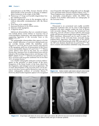
stitute radiographic evidence of navicular disease. Figure 4.8. Multiple smaller cystic lesions along the distal border
However, distal border fragments appear to be recognized of the navicular bone as demonstrated on an oblique radiograph.
A B
Figure 4.7. Single large cystic lesion (A; arrow) and multiple cystic lesions (B) within the navicular bone demonstrated on oblique
radiographs of two different horses with signs of navicular syndrome.