
Page 471 - Adams and Stashak's Lameness in Horses, 7th Edition
P. 471
Diagnostic Imaging 437
95.0 °F 98.0 °F
A B
VetBooks.ir 90 95
90
85
85
80
80
75.0 78.0
98.0 °F 100.0 °F
C D
95
95
90
90
85
85
80
78.0 80.0
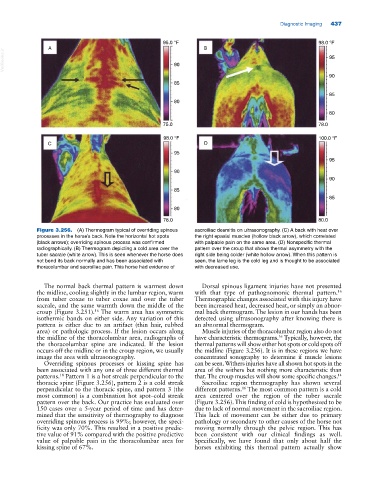
Figure 3.256. (A) Thermogram typical of overriding spinous sacroiliac desmitis on ultrasonography. (C) A back with heat over
processes in the horse’s back. Note the horizontal hot spots the right epaxial muscles (hollow black arrow), which correlated
(black arrows); overriding spinous process was confirmed with palpable pain on the same area. (D) Nonspecific thermal
radiographically. (B) Thermogram depicting a cold area over the pattern over the croup that shows thermal asymmetry with the
tuber sacrale (white arrow). This is seen whenever the horse does right side being colder (white hollow arrow). When this pattern is
not bend its back normally and has been associated with seen, the lame leg is the cold leg and is thought to be associated
thoracolumbar and sacroiliac pain. This horse had evidence of with decreased use.
The normal back thermal pattern is warmest down Dorsal spinous ligament injuries have not presented
16
the midline, cooling slightly in the lumbar region, warm with that type of pathognomonic thermal pattern.
from tuber coxae to tuber coxae and over the tuber Thermographic changes associated with this injury have
sacrale, and the same warmth down the middle of the been increased heat, decreased heat, or simply an abnor
croup (Figure 3.251). The warm area has symmetric mal back thermogram. The lesion in our hands has been
16
isothermic bands on either side. Any variation of this detected using ultrasonography after knowing there is
pattern is either due to an artifact (thin hair, rubbed an abnormal thermogram.
area) or pathologic process. If the lesion occurs along Muscle injuries of the thoracolumbar region also do not
the midline of the thoracolumbar area, radiographs of have characteristic thermograms. Typically, however, the
16
the thoracolumbar spine are indicated. If the lesion thermal patterns will show either hot spots or cold spots off
occurs off the midline or in the croup region, we usually the midline (Figure 3.256). It is in these regions we have
image the area with ultrasonography. concentrated sonography to determine if muscle lesions
Overriding spinous processes or kissing spine has can be seen. Withers injuries have all shown hot spots in the
been associated with any one of three different thermal area of the withers but nothing more characteristic than
patterns. Pattern 1 is a hot streak perpendicular to the that. The croup muscles will show some specific changes. 14
18
thoracic spine (Figure 3.256), pattern 2 is a cold streak Sacroiliac region thermography has shown several
perpendicular to the thoracic spine, and pattern 3 (the different patterns. The most common pattern is a cold
16
most common) is a combination hot spot–cold streak area centered over the region of the tuber sacrale
pattern over the back. Our practice has evaluated over (Figure 3.256). This finding of cold is hypothesized to be
150 cases over a 5‐year period of time and has deter due to lack of normal movement in the sacroiliac region.
mined that the sensitivity of thermography to diagnose This lack of movement can be either due to primary
overriding spinous process is 99%; however, the speci pathology or secondary to other causes of the horse not
ficity was only 70%. This resulted in a positive predic moving normally through the pelvic region. This has
tive value of 91% compared with the positive predictive been consistent with our clinical findings as well.
value of palpable pain in the thoracolumbar area for Specifically, we have found that only about half the
kissing spine of 67%. horses exhibiting this thermal pattern actually show