
Page 465 - Adams and Stashak's Lameness in Horses, 7th Edition
P. 465
Diagnostic Imaging 431
THERMOGRAPHY
VetBooks.ir tracy a. turner
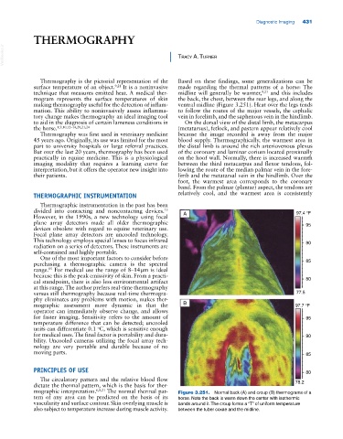
Thermography is the pictorial representation of the Based on these findings, some generalizations can be
surface temperature of an object. 9,21 It is a noninvasive made regarding the thermal patterns of a horse: The
technique that measures emitted heat. A medical ther midline will generally be warmer, 9,21 and this includes
mogram represents the surface temperatures of skin the back, the chest, between the rear legs, and along the
making thermography useful for the detection of inflam ventral midline (Figure 3.251). Heat over the legs tends
mation. This ability to noninvasively assess inflamma to follow the routes of the major vessels, the cephalic
tory change makes thermography an ideal imaging tool vein in forelimb, and the saphenous vein in the hindlimb.
to aid in the diagnosis of certain lameness conditions in On the dorsal view of the distal limb, the metacarpus
the horse. 1,9,10,12–14,20,21,24 (metatarsus), fetlock, and pastern appear relatively cool
Thermography was first used in veterinary medicine because the image recorded is away from the major
45 years ago. Originally, its use was limited for the most blood supply. Thermographically, the warmest area in
part to university hospitals or large referral practices. the distal limb is around the rich arteriovenous plexus
But over the last 20 years, thermography has been used of the coronary and laminar corium located proximally
practically in equine medicine. This is a physiological on the hoof wall. Normally, there is increased warmth
imaging modality that requires a learning curve for between the third metacarpus and flexor tendons, fol
interpretation, but it offers the operator new insight into lowing the route of the median palmar vein in the fore
their patients. limb and the metatarsal vein in the hindlimb. Over the
foot, the warmest area corresponds to the coronary
band. From the palmar (plantar) aspect, the tendons are
THERMOGRAPHIC INSTRUMENTATION relatively cool, and the warmest area is consistently
Thermographic instrumentation in the past has been
15
divided into contacting and noncontacting devices. A 97.4 °F
However, in the 1990s, a new technology using focal
plane array detectors made all older thermographic 95
devices obsolete with regard to equine veterinary use.
Focal plane array detectors are uncooled technology.
This technology employs special lenses to focus infrared 90
radiation on a series of detectors. These instruments are
self‐contained and highly portable.
One of the most important factors to consider before
purchasing a thermographic camera is the spectral 85
range. For medical use the range of 8–14 μm is ideal
15
because this is the peak emissivity of skin. From a practi
cal standpoint, there is also less environmental artifact 80
at this range. The author prefers real‐time thermography
versus still thermography because real‐time thermogra 77.5
phy eliminates any problems with motion, makes ther
mographic assessment more dynamic in that the B 97.7 °F
operator can immediately observe change, and allows
for faster imaging. Sensitivity refers to the amount of 95
temperature difference that can be detected; uncooled
units can differentiate 0.1 C, which is sensitive enough
o
for medical uses. The final factor is portability and dura 90
bility. Uncooled cameras utilizing the focal array tech
nology are very portable and durable because of no
moving parts. 85
PRINCIPLES OF USE 80
The circulatory pattern and the relative blood flow
dictate the thermal pattern, which is the basis for ther 78.2
mographic interpretation. 6,8,21 The normal thermal pat Figure 3.251. Normal back (A) and croup (B) thermograms of a
tern of any area can be predicted on the basis of its horse. Note the back is warm down the center with isothermic
vascularity and surface contour. Skin overlying muscle is bands around it. The croup forms a “T” of uniform temperature
also subject to temperature increase during muscle activity. between the tuber coxae and the midline.