
Page 498 - Adams and Stashak's Lameness in Horses, 7th Edition
P. 498
464 Chapter 4
can improve the specificity of the block for the joint. 17,55
Most horses with DIP joint pain improve rapidly and
VetBooks.ir partially improves but persists after 10 minutes, the DIP
substantially after IA anesthesia. If the lameness only
joint is not likely the primary site of the pain. In addi-
tion, a positive response to an IA block combined with a
negative response to navicular bursa anesthesia often
55
incriminates the joint as the primary problem area. See
Chapter 2 for more details related to perineural and
intrasynovial anesthesia.
Diagnosis
A definitive diagnosis of OA of the DIP joint can
often be obtained with radiography of the foot. However,
horses with synovitis/capsulitis or early OA of the DIP
joint may have no radiographic abnormalities. A com-
plete radiographic study of the foot should be performed
to rule out other potential problems because DIP joint
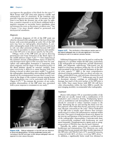
OA may be secondary to other conditions and anesthe- Figure 4.27. The calcification of the extensor tendon seen on
sia of the DIP joint is not always specific for the joint. this lateral radiograph was not clinically significant in this horse
Oblique views of the DIP joint can aid in detecting following removal of an extensor process fracture.
periarticular new bone formation of the distal aspect of
the middle phalanx (Figure 4.26). 17,39 Close inspection of
the extensor process, palmar/plantar aspect of distal P2, Additional diagnostics that may be used to confirm the
and dorsoproximal aspect of the navicular bone for oste- diagnosis of a problem within the DIP joint, particularly
ophyte and enthesophyte formation is important. Joint if the radiographs are nondiagnostic, include ultrasound,
space congruity and the shape of the proximal surface of MRI, and diagnostic arthroscopy. Ultrasound can be
the distal phalanx should be assessed carefully. Joint helpful to document problems within the CLs of the DIP
space narrowing is difficult to assess because limb posi- joint proximal to the hoof wall and thickness of the dor-
tioning is known to affect joint space width. In general, sal joint capsule. 12,19 MRI is the most comprehensive
10
the radiographic abnormalities surrounding the DIP joint advanced imaging modality that can detect articular car-
should not be overinterpreted because there is much vari- tilage, subchondral bone, and soft tissue abnormalities of
ation in the shape of the extensor process among horses the DIP joint if present. 18,20,21,26 Arthroscopy may be used
and enthesophytes may not be associated with lameness to document articular cartilage or subchondral bone
(Figure 4.27). 17,47 In contrast, the presence of any radio- damage, but much of the joint surface of the DIP joint is
graphic abnormalities of the DIP joint was associated not visible. Since many horses with DIP OA will have
with a poor response to treatment in one study. 16 other abnormalities within the foot, MRI is usually the
next imaging modality recommended after radiography.
Treatment
Horses with primary OA or synovitis/capsulitis of the
DIP joint are usually treated with a combination of IA
medication and corrective shoeing. Predisposing factors
such as mediolateral and dorsopalmar hoof imbalances
should be corrected to reduce repetitive trauma to the
joint. Shortening the toe and moving the break‐over fur-
ther palmarly often helps these horses, and using a rim pad
may alleviate concussion to the joint. Direct medication
5
of the DIP joint is usually more effective than systemic
medications to reduce the inflammatory response within
the joint. The type(s) of medication chosen may depend on
the severity and duration of the lameness and the severity
of the radiographic abnormalities. Corticosteroids alone
or corticosteroids combined with hyaluronan are used
most frequently. Chondroprotective agents such as autog-
enous serum or PSGAGs may also be used if desired.
Typically, the author recommends a combination of
hyaluronan and triamcinolone (TA). However, one study
Figure 4.26. Oblique radiographs of the DIP joint are important reported that horses treated with three weekly injections of
to document abnormalities. The bony proliferation (arrow) and the DIP joint with PSGAG had a 67% successful outcome,
35
narrowing of the DIP joint seen on this oblique radiograph were not compared with 46% of those receiving MPA alone.
apparent on other views of the joint. In addition, a significantly better result was obtained in