
Page 499 - Adams and Stashak's Lameness in Horses, 7th Edition
P. 499
Lameness of the Distal Limb 465
dressage horses than in jumping horses (eventing and show identified 65 cases of P3 fractures in 20,638 cases admitted
56
35
jumping). This study and the clinical observations of the to a hospital. These fractures can occur in any foot but
VetBooks.ir joint pain with IA medication although horses that block limb and the medial aspect of the right forelimb in race-
most commonly affect the lateral aspect of the left fore-
author support the rationale for treating horses with DIP
Type I and II “wing” fractures are most
completely with DIP joint anesthesia usually respond the
horses.
5,47
best to this treatment. Repeat IA injections may be required, common and the majority of these fractures enter the
depending on the severity of the abnormalities within the DIP joint. 47,56 In one report of P3 fractures in
joint and the response to treatment. Thoroughbred and Standardbred racehorses, 71 of 74
Treatment of horses with secondary OA of the DIP fractures were wing fractures (types I and II), and the
47
joint usually focuses on the underlying contributing majority of these fractures were articular. Although all
problem. Treatment of the primary condition is usually breeds and classes of horses can be affected, there appears
beneficial to prevent worsening of the problems within to be a higher incidence observed in racing breeds. 5,47,56
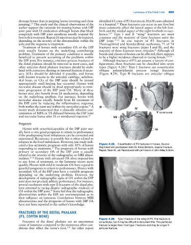
the DIP joint. For instance, extensor process fractures of Although fractures of P3 can assume a variety of con-
the distal phalanx should be removed in most cases, and figurations, these fractures can be classified into seven
other articular distal phalanx fractures should be stabi- types (Figure 4.28). Type I fractures are nonarticular
6
lized with corrective shoeing or internal fixation if neces- oblique palmar/plantar process (wing) fractures
sary. SCLs should be debrided if possible, and horses (Figure 4.29). Type II fractures are articular oblique
with known trauma to the articular cartilage, subchon-
dral bone, or CLs of the DIP joint should be treated
appropriately until healing has occurred. Horses with
navicular disease should be shod appropriately to mini- I
mize progression of the DIP joint OA. Many of these
horses may also benefit from IA medication, depending
on the underlying problem. For instance, horses with
navicular disease usually benefit from IA treatment of II
the DIP joint by reducing the inflammatory response,
both within the joint and within the navicular region. A III
45
recent study documented that a clinically effective con- IV
centration of MPA or TA diffused between the DIP joint V (comminuted)
and navicular bursa after IA or intrabursal injection. 45 VII
Prognosis
Horses with synovitis/capsulitis of the DIP joint usu- VI
ally have a very good prognosis to return to performance
if the predisposing hoof imbalances can be corrected and
maintained. Recurrence is possible but is often related to
relapses in the hoof imbalances. However, one study indi-
cated a less optimistic prognosis with only 30% of horses Figure 4.28. Classification of P3 fractures in horses. Source:
16
responding to treatment. The prognosis of horses with Reprinted with permission from Dr. Alicia Bertone, Equine Fracture
primary or secondary OA of the DIP joint is usually Repair, Nixon AL, ed. Reproduced with permission of John Wiley & Sons.
related to the severity of the radiographic or MRI abnor-
malities. 17,18 Horses with advanced OA often respond less
to any form of treatment, or the lameness recurs more
quickly. Horses with mild to moderate OA have a good to
guarded prognosis to return to performance. Horses with
secondary OA of the DIP joint have a variable prognosis
depending on the underlying problem. However, the
development of radiographic signs of OA within the DIP
joint does not preclude athletic performance. For instance,
several racehorses with type II fractures of the distal pha-
lanx returned to racing despite radiographic evidence of
OA within the DIP joint. Some feel that the radiographic
47
abnormalities within the DIP are overinterpreted as to
their influence on lameness. Correlations between MRI
abnormalities and the prognosis of horses with DIP OA
have not been reported to the author’s knowledge.
FRACTURES OF THE DISTAL PHALANX
(P3, COFFIN BONE)
Figure 4.29. Type I fracture of the wing of P3. This fracture is
Fractures of the distal phalanx are an uncommon nonarticular, but it may be difficult to document this. This particular
cause of lameness compared to the numerous other con- fracture is larger than most type I fractures and may be a type II
ditions that affect the horse’s foot. 5,56 An older report articular fracture.