
Page 500 - Adams and Stashak's Lameness in Horses, 7th Edition
P. 500
466 Chapter 4
palmar or plantar process (wing) fractures (Figure 4.30) triangular or oblong in shape. 30,32 Initially they were
and are by far the most common type. 47,56 Type III frac- thought to represent osseous bodies, but histologic find-
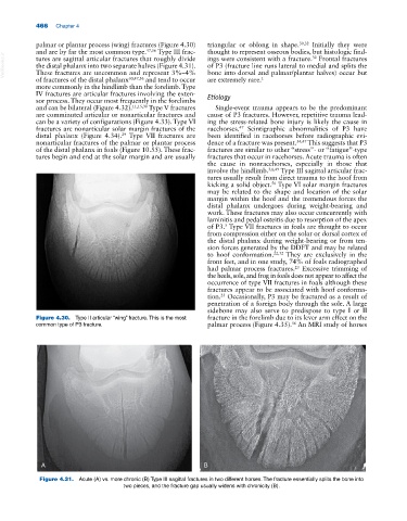
VetBooks.ir the distal phalanx into two separate halves (Figure 4.31). of P3 (fracture line runs lateral to medial and splits the
ings were consistent with a fracture. Frontal fractures
tures are sagittal articular fractures that roughly divide
32
These fractures are uncommon and represent 3%–4%
bone into dorsal and palmar/plantar halves) occur but
of fractures of the distal phalanx 30,47,56 and tend to occur are extremely rare. 1
more commonly in the hindlimb than the forelimb. Type
IV fractures are articular fractures involving the exten- Etiology
sor process. They occur most frequently in the forelimbs
and can be bilateral (Figure 4.32). 11,15,30 Type V fractures Single‐event trauma appears to be the predominant
are comminuted articular or nonarticular fractures and cause of P3 fractures. However, repetitive trauma lead-
can be a variety of configurations (Figure 4.33). Type VI ing the stress‐related bone injury is likely the cause in
fractures are nonarticular solar margin fractures of the racehorses. Scintigraphic abnormalities of P3 have
47
29
distal phalanx (Figure 4.34). Type VII fractures are been identified in racehorses before radiographic evi-
nonarticular fractures of the palmar or plantar process dence of a fracture was present. 34,47 This suggests that P3
of the distal phalanx in foals (Figure 10.55). These frac- fractures are similar to other “stress”‐ or “fatigue”‐type
tures begin and end at the solar margin and are usually fractures that occur in racehorses. Acute trauma is often
the cause in nonracehorses, especially in those that
involve the hindlimb. 3,6,49 Type III sagittal articular frac-
tures usually result from direct trauma to the hoof from
kicking a solid object. Type VI solar margin fractures
56
may be related to the shape and location of the solar
margin within the hoof and the tremendous forces the
distal phalanx undergoes during weight‐bearing and
work. These fractures may also occur concurrently with
laminitis and pedal osteitis due to resorption of the apex
of P3. Type VII fractures in foals are thought to occur
5
from compression either on the solar or dorsal cortex of
the distal phalanx during weight‐bearing or from ten-
sion forces generated by the DDFT and may be related
to hoof conformation. 22,32 They are exclusively in the
front feet, and in one study, 74% of foals radiographed
had palmar process fractures. Excessive trimming of
23
the heels, sole, and frog in foals does not appear to affect the
occurrence of type VII fractures in foals although these
fractures appear to be associated with hoof conforma-
tion. Occasionally, P3 may be fractured as a result of
23
penetration of a foreign body through the sole. A large
sidebone may also serve to predispose to type I or II
Figure 4.30. Type II articular “wing” fracture. This is the most fracture in the forelimb due to its lever arm effect on the
56
common type of P3 fracture. palmar process (Figure 4.35). An MRI study of horses
A B
Figure 4.31. Acute (A) vs. more chronic (B) Type III sagittal fractures in two different horses. The fracture essentially splits the bone into
two pieces, and the fracture gap usually widens with chronicity (B).