
Page 502 - Adams and Stashak's Lameness in Horses, 7th Edition
P. 502
468 Chapter 4
surface. Smaller type IV fractures rarely cause deformity
of the dorsal hoof wall, but effusion of the DIP joint is
VetBooks.ir Diagnosis
common.
Radiographic examination (30° dorsopalmar/plantar,
65° dorsoproximal to palmarodistal, lateral, and both
obliques) is used to confirm the diagnosis and document
the type and location of the fracture (see Chapter 3 for
more details on radiographic views). In some cases it
may be necessary to take special views of the palmar/
plantar processes to identify the fracture. Solar margin
fractures are most easily identified on the 60° dorso-
proximal to palmarodistal projection using a radio-
graphic technique with approximately one‐half the
exposure needed to evaluate the navicular bone.
30
Extensor process fractures are usually identified on the
lateromedial view.
Most P3 fractures are readily apparent on routine
radiographic projections. However, nondisplaced or
stress‐related fractures in racehorses may not be appar-
ent on the initial radiographic examination because of
insufficient time for resorption of the bone along the
fracture line or because the cast‐like effect of the hoof
wall may prevent fracture displacement. 30,34 In these
cases, radiographs should be repeated in 1–2 weeks, or
nuclear scintigraphy, CT, or MRI can be used to help
identify radiographically occult fractures of P3. 5,37,47 In
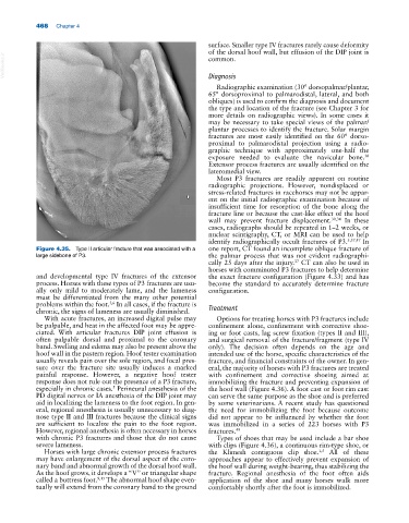
Figure 4.35. Type II articular fracture that was associated with a one report, CT found an incomplete oblique fracture of
large sidebone of P3. the palmar process that was not evident radiographi-
37
cally 25 days after the injury. CT can also be used in
horses with comminuted P3 fractures to help determine
and developmental type IV fractures of the extensor the exact fracture configuration (Figure 4.33) and has
process. Horses with these types of P3 fractures are usu- become the standard to accurately determine fracture
ally only mild to moderately lame, and the lameness configuration.
must be differentiated from the many other potential
problems within the foot. In all cases, if the fracture is
5,6
chronic, the signs of lameness are usually diminished. Treatment
With acute fractures, an increased digital pulse may Options for treating horses with P3 fractures include
be palpable, and heat in the affected foot may be appre- confinement alone, confinement with corrective shoe-
ciated. With articular fractures DIP joint effusion is ing or foot casts, lag screw fixation (types II and III),
often palpable dorsal and proximal to the coronary and surgical removal of the fracture/fragment (type IV
band. Swelling and edema may also be present above the only). The decision often depends on the age and
hoof wall in the pastern region. Hoof tester examination intended use of the horse, specific characteristics of the
usually reveals pain over the sole region, and focal pres- fracture, and financial constraints of the owner. In gen-
sure over the fracture site usually induces a marked eral, the majority of horses with P3 fractures are treated
painful response. However, a negative hoof tester with confinement and corrective shoeing aimed at
response does not rule out the presence of a P3 fracture, immobilizing the fracture and preventing expansion of
especially in chronic cases. Perineural anesthesia of the the hoof wall (Figure 4.36). A foot cast or foot rim cast
5
PD digital nerves or IA anesthesia of the DIP joint may can serve the same purpose as the shoe and is preferred
aid in localizing the lameness to the foot region. In gen- by some veterinarians. A recent study has questioned
eral, regional anesthesia is usually unnecessary to diag- the need for immobilizing the foot because outcome
nose type II and III fractures because the clinical signs did not appear to be influenced by whether the foot
are sufficient to localize the pain to the foot region. was immobilized in a series of 223 horses with P3
However, regional anesthesia is often necessary in horses fractures. 49
with chronic P3 fractures and those that do not cause Types of shoes that may be used include a bar shoe
severe lameness. with clips (Figure 4.36), a continuous rim‐type shoe, or
Horses with large chronic extensor process fractures the Klimesh contiguous clip shoe. All of these
1,5
may have enlargement of the dorsal aspect of the coro- approaches appear to effectively prevent expansion of
nary band and abnormal growth of the dorsal hoof wall. the hoof wall during weight‐bearing, thus stabilizing the
As the hoof grows, it develops a “V” or triangular shape fracture. Regional anesthesia of the foot often aids
called a buttress foot. 9,15 The abnormal hoof shape even- application of the shoe and many horses walk more
tually will extend from the coronary band to the ground comfortably shortly after the foot is immobilized.