
Page 507 - Adams and Stashak's Lameness in Horses, 7th Edition
P. 507
Lameness of the Distal Limb 473
to the middle phalanx, contributing to injuries to the CL lame horses reported a correlation between a reduced
of the DIP joint. This can occur as the result of an acute palmar angle of the foot detected with radiography and
VetBooks.ir within the CL of the DIP joint have recently been
alterations in the CLs of the DIP joint with MRI.
injury or from repetitive‐type trauma. However, lesions
14
reported to be a primary degenerative process rather
20
than inflammatory. Histology revealed extensive fibro- Clinical Signs
cartilaginous metaplasia and development of multiple In general, horses that have primary soft tissue injuries
intercommunicating fissures within the degenerate col- in the foot such as injuries to the CLs of the DIP joint are
lagen in severe lesions. This was thought to explain the more likely to have a history of an acute onset of lame-
20
poor response to conservative treatment in many horses ness and be unilaterally lame compared with horses with
with desmitis of the CL of the DIP joint. There are no injuries to the navicular region. There are often few
known predisposing factors, but a recent study of 52 localizing clinical signs in horses with injuries to the CL
of the DIP joint.19, 26 Horses often have a history of a
chronic forelimb lameness of variable severity that is
worse in the circle. Palpable swelling and pain of the CL
at its proximal attachment to the middle phalanx may be
present above the coronary band in severe cases.
However, this is uncommon and effusion of the DIP joint
is also not a consistent clinical finding. Most horses
(87% in one study) improve with a PD nerve block but
may not be completely sound until a more proximal
block is performed.19 Only 40% of horses improved
with IA anesthesia of the DIP joint in one study.19
Diagnosis
A definitive diagnosis of an injury to the CL of the
DIP joint is best determined with MRI in either the
recumbent or standing patient. Lesions within the CL of
the DIP joint are identified by the alteration in size and
signal intensity (Figure 4.42). In addition, some horses
may have abnormal mineralization and fluid within the
distal phalanx at the insertion of the ligament (see
18
Figure 4.41. Bony proliferation on the dorsolateral aspect of P2 Chapter 3 for more information on MRI). Ultrasound
visible on this oblique radiograph (arrow) may be suggestive of a may also be helpful, but certain aspects of the CL of the
chronic injury to the CL of the DIP joint. DIP joint are inaccessible with ultrasound, suggesting
A B
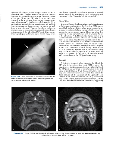
Figure 4.42. Frontal STIR (A) and PD axial (B) MR images of the foot of a 12‐year‐old Quarter horse with abnormalities within the
medial collateral ligament of the DIP joint (arrows).