
Page 509 - Adams and Stashak's Lameness in Horses, 7th Edition
P. 509
Lameness of the Distal Limb 475
from the junction between a separate ossification center
and that part of the cartilage that is ossifying from the
VetBooks.ir ossified cartilage can occur but is rare.
A fracture in the
palmar process of the distal phalanx.
5,51
Documenting that sidebone is the cause of lameness in
horses can be difficult. Asymmetrical swelling of pastern
region, pain on palpation of the collateral cartilage, and
improvement of the lameness with a uniaxial PD nerve
block are suggestive of a problem in this region. However,
the mere presence of an ossified cartilage without any
other radiographic abnormalities is not diagnostic. The
clinical relevance of these findings should be further doc-
umented with scintigraphy and/or MRI. 41,42
Treatment
If sidebone is suspected as the cause of lameness, con-
servative treatment with rest, topical 1% diclofenac
sodium cream (Surpass®), and oral administration of
NSAIDs is recommended initially. External compression
of the site together with cold‐water therapy may also be
beneficial. Any contributing foot problems such as foot
imbalances should be addressed. Surgical removal of sus-
pected fractured sidebones is not recommended. If lame-
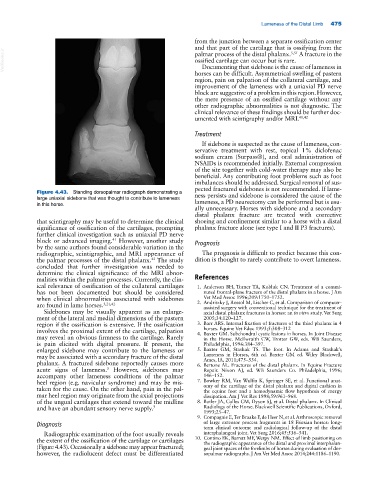
Figure 4.43. Standing dorsopalmar radiograph demonstrating a ness persists and sidebone is considered the cause of the
large uniaxial sidebone that was thought to contribute to lameness lameness, a PD neurectomy can be performed but is usu-
in this horse.
ally unnecessary. Horses with sidebone and a secondary
distal phalanx fracture are treated with corrective
that scintigraphy may be useful to determine the clinical shoeing and confinement similar to a horse with a distal
significance of ossification of the cartilages, prompting phalanx fracture alone (see type I and II P3 fractures).
further clinical investigation such as uniaxial PD nerve
block or advanced imaging. However, another study Prognosis
41
by the same authors found considerable variation in the
radiographic, scintigraphic, and MRI appearance of The prognosis is difficult to predict because this con-
the palmar processes of the distal phalanx. The study dition is thought to rarely contribute to overt lameness.
42
concluded that further investigation was needed to
determine the clinical significance of the MRI abnor-
malities within the palmar processes. Currently, the clin- References
ical relevance of ossification of the collateral cartilages 1. Anderson BH, Turner TA, Kobluk CN. Treatment of a commi-
has not been documented but should be considered nuted frontal‐plane fracture of the distal phalanx in a horse. J Am
when clinical abnormalities associated with sidebones Vet Med Assoc 1996;209:1750–1752.
are found in lame horses. 5,21,42 2. Andritzky J, Rossol M, Lischer C, et al. Comparison of computer‐
assisted surgery with conventional technique for the treatment of
Sidebones may be visually apparent as an enlarge- axial distal phalanx fractures in horses: an in vitro study. Vet Surg
ment of the lateral and medial dimensions of the pastern 2005;34:120–127.
region if the ossification is extensive. If the ossification 3. Barr ARS. Internal fixation of fractures of the third phalanx in 4
horses. Equine Vet Educ 1993;5:308–312
involves the proximal extent of the cartilage, palpation 4. Baxter GM. Subchondral cystic lesions in horses. In Joint Disease
may reveal an obvious firmness to the cartilage. Rarely in the Horse. McIlwraith CW, Trotter GW, eds. WB Saunders,
is pain elicited with digital pressure. If present, the Philadelphia, 1996:384–397.
enlarged sidebone may contribute to the lameness or 5. Baxter GM, Stashak TS. The foot. In Adams and Stashak’s
may be associated with a secondary fracture of the distal Lameness in Horses, 6th ed. Baxter GM ed. Wiley Blackwell,
Ames, IA, 2011;475–534.
phalanx. A fractured sidebone reportedly causes more 6. Bertone AL. Fractures of the distal phalanx. In Equine Fracture
acute signs of lameness. However, sidebones may Repair. Nixon AJ, ed. WB Saunders Co. Philadelphia, 1996;
5
accompany other lameness conditions of the palmar 146–152.
heel region (e.g. navicular syndrome) and may be mis- 7. Bowker RM, Van Wulfin K, Springer SE, et al. Functional anat-
taken for the cause. On the other hand, pain in the pal- omy of the cartilage of the distal phalanx and digital cushion in
the equine foot and a hemodynamic flow hypothesis of energy
mar heel region may originate from the axial projections dissipation. Am J Vet Res 1998;59:961–968.
of the ungual cartilages that extend toward the midline 8. Butler JA, Colles CM, Dyson SJ, et al. Distal phalanx. In Clinical
and have an abundant sensory nerve supply. 7 Radiology of the Horse. Blackwell Scientific Publications, Oxford,
1993;25–47.
9. Compagnie E, Ter Braake F, de Heer N, et al. Arthroscopic removal
Diagnosis of large extensor process fragments in 18 Friesian horses: long‐
term clinical outcome and radiological follow‐up of the distal
Radiographic examination of the foot usually reveals interphalangeal joint. Vet Surg 2016;45:536–541.
the extent of the ossification of the cartilage or cartilages 10. Contino EK, Barrett MF, Werpy NM. Effect of limb positioning on
the radiographic appearance of the distal and proximal interphalan-
(Figure 4.43). Occasionally a sidebone may appear fractured; geal joint spaces of the forelimbs of horses during evaluation of dor-
however, the radiolucent defect must be differentiated sopalmar radiographs. J Am Vet Med Assoc 2014;244:1186–1190.