
Page 513 - Adams and Stashak's Lameness in Horses, 7th Edition
P. 513
Lameness of the Distal Limb 479
In cases in which shoeing contributes to the bruising, Etiology
removal of the shoe may be all that is necessary. The horse Environmental microbes gain access to the distal pha-
VetBooks.ir disappear. If the horse must be used, the wall and bar in lanx either by direct introduction of bacteria by deep
should be rested and should not be reshod until symptoms
penetrating injuries, extension of infection from the soft
the region of the corn should be removed to prevent pres-
sure by the shoe. A complete support shoe can be applied tissues of the foot into the distal phalanx, or from hema-
23,31
to allow the frog to absorb the concussion that would nor- togenous sources in foals. Septic PO in foals should
mally be distributed to the corn area. Another option is to be considered as a potential site for hematogenous
apply a wide‐web shoe that has been concaved out at the spread of infection associated with the septic arthritis/
24
damaged region to decrease the pressure on the bruised joint ill syndrome. One study found that 8 of 22 foals
site. To prevent shoes from causing corns, the heels of the with septic PO had multiple other foci of infection and
shoes should extend well back on the buttresses and none of the foals had a history of a penetrating wound
23
should fit full on the wall at the quarters and heels. 31 or subsolar abscess. Other causes of septic PO in adult
horses include chronic severe laminitis, subsolar
abscesses (most common), solar margin fractures, deep
Prognosis hoof wall cracks, avulsion hoof injuries, and penetrating
The prognosis is usually very good for horses suffer- wounds of the foot. 6,11,19 A sequestrum may develop in
6,16
ing from a single traumatic episode and in those with the distal phalanx as the osseous infection progresses.
good foot conformation. The prognosis is reduced in In a review of 63 horses treated for septic PO, subsolar
horses with poor hoof conformation that are continually abscesses were the most common cause (56%) followed
worked on hard ground because recurrence is common. by solar margin fractures (25%) and penetrating wounds
19
Many of these cases develop chronic sole bruising, which (13%).
eventually may lead to chronic abnormalities within the
distal phalanx. Horses with routine foot abscesses also Clinical Signs
have a very good prognosis, provided the infection does
not involve deeper structures in the foot. It should always Septic PO occurs most commonly in the forelimbs in
be remembered that subsolar abscesses may be associ- adult horses and in the hindlimbs in foals. 6,23 The fore-
ated with other conditions of the foot such as keratomas, limbs were affected twice as commonly as the hindlimbs
7
infection of the collateral cartilages, chronic laminitis, in one study, and 11 of 18 cases involved the forelimbs
6
and septic pedal osteitis (PO) (Figure 4.47). in another study. The severity of lameness in horses
with septic PO is usually severe (in one study over 50%
of the horses were grade 4 of 5 lame), and the lameness
SEPTIC PEDAL OSTEITIS may be chronic (average duration of lameness prior to
presentation in one study was 18.5 days). 6,19 Increased
Septic PO refers to bacterial infection within the distal hoof temperature and prominent digital pulses are com-
phalanx. It can occur as an extension of a subsolar mon in the affected limb. Hoof tester exam may be ben-
abscess, secondary to penetrating injuries or from hema- eficial to localize the site of pain as well as to promote
togenous spread in foals. Radiographically the condition abscess drainage in some cases. Perineural anesthesia of
usually appears as a focal or diffuse radiolucency of the the PD nerves may not eliminate the lameness in all
bone (osteolysis). The distal phalanx does not have a horses with septic PO. No or minimal response to PD
6,9
medullary cavity, so inflammation/infection of this bone nerve block was seen in 5 of 5 cases of septic PO, and all
is referred to as osteitis rather than osteomyelitis. 11 5 cases improved after an abaxial sesamoid nerve block. 6
Diagnosis
Radiographic signs suggestive of septic PO are usu-
ally straightforward and are consistent with areas of
bone infection (Figure 4.48). CT examination of the
foot can often provide more detailed information but is
usually unnecessary to make a diagnosis. Generally,
there is a loss of trabecular detail (osteolysis) with indis-
tinct margins fading into the surrounding bone.
31
Sequestra may develop but marginal sclerosis is rarely
observed in these cases (Figure 4.49). Radiographic
19
examination of 18 horses diagnosed with septic PO
revealed discrete osteolysis at the margins of the distal
phalanx (13 of 18 cases), gas density adjacent to the
bone on 2 different radiographic projections (15 of 15
cases), focal (9 cases) or diffuse (3 cases) decrease in
bone density, generalized roughening of the solar margin
of the distal phalanx (7 of 18 cases), and widening of the
6
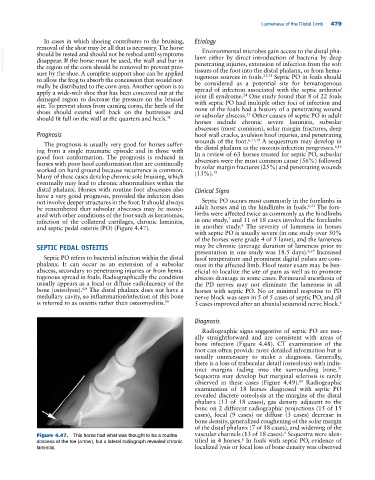
Figure 4.47. This horse had what was thought to be a routine vascular channels (13 of 18 cases). Sequestra were iden-
6
abscess at the toe (arrow), but a lateral radiograph revealed chronic tified in 4 horses. In foals with septic PO, evidence of
laminitis. localized lysis or focal loss of bone density was observed