
Page 515 - Adams and Stashak's Lameness in Horses, 7th Edition
P. 515
Lameness of the Distal Limb 481
Of 33 cases of septic PO in which follow‐up was avail-
19
able, all 33 horses returned to their intended use. In
VetBooks.ir use, and 13 of 18 had a favorable outcome despite a
other studies, 7 of 9 horses returned to their intended
11
high incidence of sequestra and fractures. Foals with
6
septic PO also have a good prognosis; 86% of the foals
survived, 16 of 22 foals reached racing age, and 11 of
these raced. 23,24
PENETRATING INJURIES OF THE FOOT
Penetrating injuries of the foot are commonly seen in
equine practice. The injury is often sustained by the
horse stepping on (bottom of the foot) or contacting
(coronary band, heel bulbs, and pastern region) a sharp
object. Although any deep penetrating injury to the foot
can potentially be serious, those that penetrate the cen-
tral third of the frog, the coronary band, and the heel
bulbs are at greatest risk to involve deeper vital struc-
tures. Structures at risk with injuries to the central third
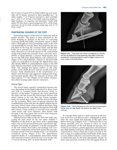
of the frog include the navicular bursa, navicular bone, Figure 4.51. This horse had a history of stepping on a farriery
DDFT, or distal phalanx. Coronary band injuries may nail with its hindfoot 24 hours previously. Exploration of the tract
involve the DIP joint, distal phalanx, and collateral car- revealed that it had penetrated through the digital cushion to the
tilages of the distal phalanx. Injuries to the heel bulb solar surface of the third phalanx.
region are most likely to damage the digital flexor ten-
don sheath, DDFT, or palmar/plantar aspect of the DIP
joint. Injuries elsewhere on the bottom of the foot most
likely only damage the digital cushion or the solar
surface of the distal phalanx and are usually less prob-
lematic. Early identification of the involvement of a
deeper vital structure and aggressive medical and surgi-
cal treatment greatly affect the outcome of horses with
these injuries. 2,3,9,29 See Chapter 12 for further informa-
tion about treating septic synovial structures.
Clinical Signs
The clinical signs caused by penetrating injuries may
vary depending on the depth (superficial vs. deep), loca-
tion (sole vs. coronary band), and duration (acute vs.
3
chronic) of the injury. In general, the more superficial
the injury, the less severe the clinical signs. Horses with
minor injuries to the sole that may remain asympto-
matic for several days until a subsolar abscess develops
are the exception. These types of injuries penetrate the
cornified layer of the sole into the digital cushion but do
not cause clinical signs unless an abscess develops. Deep
injuries that penetrate completely through the hoof wall Figure 4.52. Taking radiographs of the foot prior to removing the
and contact a bone, tendon, or synovial cavity typically foreign body can help identify the direction and depth of the
puncture.
cause severe and acute lameness. Also, horses with
wounds in the frog region that involve vital structures
usually become rapidly symptomatic. If a foreign body such as a nail is present in the bot-
Affected horses often point the foot and resist con- tom of the foot, it is ideal to take a radiograph to deter-
tacting the heel (walk on the toe) when walked. 3,32 mine the exact depth and direction of the nail’s path
Palpation of the hoof in any horse with a penetrating before removing it (Figure 4.52). 1,29 However, this is not
injury may reveal increased heat, and a prominent digi- always possible, and care should be taken to avoid
tal pulse can usually be palpated. Careful examination deeper penetration of the nail during the process. If a
2
of the sole (visual, hoof tester, and probing) and coro- wound is not obvious, careful application of hoof testers
nary band is important. It has been stated that any pen- may help identify focal pain, which may indicate the site
etrating wound of the foot deeper than 1 cm should be of penetration. Once a focal site of pain is found in the
considered serious (Figure 4.51). The approximate sole or frog, it is explored with a hoof knife. If the injury
reported depths of a perpendicular penetration before is acute (before infection) and involves the sole, a crack
vital structures become involved were 1 cm for the sole, or small hole may be the only abnormality found. In
3
1.5 cm for the frog, and 1.2 cm for the hoof wall. 3 most cases, puncture wounds of the sole appear black at