
Page 518 - Adams and Stashak's Lameness in Horses, 7th Edition
P. 518
484 Chapter 4
examination reveals abundant keratin, squamous epi-
thelial cells, occasionally granulation tissue, and inflam-
VetBooks.ir coronary band, but it may extend to the solar surface
The growth usually begins near the
matory cells.
13,20
anywhere along the white line. A visible deviation of
31
the coronary band and/or hoof wall is often present,
and the most commonly affected areas of the foot are
20
the toe and quarter. Occasionally a keratoma may be
located at a focal site between the coronary band and
sole. Lameness and the radiographic changes are
thought to arise from the growth of the keratoma and
the subsequent pressure that is applied to the sensitive
lamina and distal phalanx. Keratomas have been
20
observed in horses ranging from 2 to 20 years of age
and should be differentiated from other growths that
can occur in the hoof such as squamous cell carcinoma,
canker, and melanoma. 20,31 In addition, multiple keratomas
may be present in the same foot, but this is uncommon. 8,17
Etiology
Trauma and chronic irritation in the form of sole
abscesses or direct hoof injuries are the cause in the
majority of cases. 20,31 However, a keratoma can develop
without a history of previous injury, and the initiating
cause often cannot be determined. 17,31
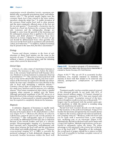
Clinical Signs Figure 4.57. Dorsopalmar radiograph of P3 demonstrating a
A history of a slow onset of intermittent lameness is smooth margined lytic defect within the bone that is characteristic of
common. The lameness is often seen before the distor- a keratoma. Source: Courtesy of Dr. Scott Katzman.
tion at the coronary band and hoof wall becomes obvi-
ous. Moderate to severe lameness is commonly observed (Figure 4.58). 12,17 The use of CT to accurately localize
at presentation. 17,20,31 The coronary band and hoof wall keratomas was recently reported to minimize the
may or may not be abnormally shaped, and close exami- amount of hoof wall that needed to be removed and
nation of the foot may be required to identify any abnor- reduced postoperative complications in operated
mality. In some cases a fistulous tract may develop in the horses. 17
sole or hoof wall, mimicking a subsolar abscess. 5,16
Common clinical signs of keratomas in one retrospec- Treatment
tive study were lameness and the presence of a subsolar
abscess. Hoof tester examination often elicits a painful Treatment usually involves complete surgical removal
5
response when pressure is applied over the lesion. of the abnormal growth. In one study only 42% of
Although perineural anesthesia of the PD nerves at or horses treated without surgery (12 horses) returned to
below the level of the collateral cartilages often improves performance compared to 83% that were treated with
4
the lameness, a basisesamoid or abaxial sesamoid block surgery (23 horses). Incomplete removal of the kera-
may be required to completely eliminate the lameness. toma is thought to result in recurrence of the growth. 17,20,31
Surgery may be performed with the patient under gen-
Diagnosis eral anesthesia or while the patient is standing using
regional anesthesia and sedation.
A definitive diagnosis of keratoma is usually made Partial hoof wall resection directly over the location
based on the characteristic radiographic features. A of the keratoma is the preferred technique. Using CT to
discrete semicircular defect in the distal phalanx is often localize the keratoma preoperatively was recently shown
seen (Figure 4.57). 5,31 However, the absence of a discrete to improve the accuracy of hoof wall removal and facili-
radiolucency cannot be used to rule out the presence of tate complete removal. Windows within the hoof wall
17
a keratoma. 17,20 The radiographic signs of a keratoma can be made with a motorized burr, a cast cutting
can usually be differentiated from lysis due to infection saw, oscillating saw, or an osteotome (Figure 4.59).
because of the smooth borders and lack of a sclerotic Alternatively, the hole in the hoof wall can be made with
margin. Ultrasonographic imaging of a keratoma has a large Galt trephine. Multiple trephinations can be per-
been reported, and a hypoechoic, well‐delineated soft formed to adequately expose the lesion. The major
tissue mass between the hoof wall and the articulation advantage to this approach is the relative lack of disrup-
of the distal and middle phalanges was seen. However, tion of the hoof wall. 16,17 Regardless of the technique,
30
cross‐sectional imaging (CT or MRI) is currently the the overall goal of surgery should be to remove as little
preferred technique to both definitively diagnose and hoof wall as possible to facilitate complete removal of
accurately localize keratomas within the hoof wall the keratoma. 5,16,17