
Page 519 - Adams and Stashak's Lameness in Horses, 7th Edition
P. 519
Lameness of the Distal Limb 485
VetBooks.ir
A B
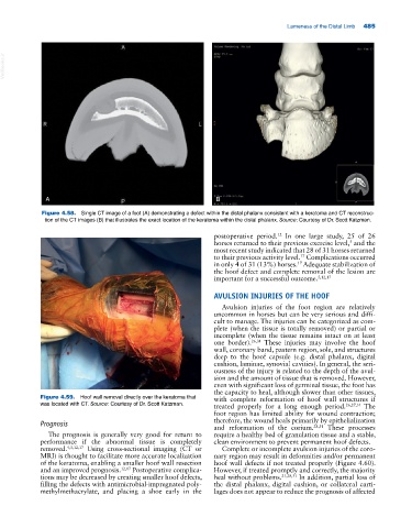
Figure 4.58. Single CT image of a foot (A) demonstrating a defect within the distal phalanx consistent with a keratoma and CT reconstruc-
tion of the CT images (B) that illustrates the exact location of the keratoma within the distal phalanx. Source: Courtesy of Dr. Scott Katzman.
postoperative period. In one large study, 25 of 26
12
horses returned to their previous exercise level, and the
5
most recent study indicated that 28 of 31 horses returned
to their previous activity level. Complications occurred
17
in only 4 of 31 (13%) horses. Adequate stabilization of
17
the hoof defect and complete removal of the lesion are
important for a successful outcome. 5,12,17
AVULSION INJURIES OF THE HOOF
Avulsion injuries of the foot region are relatively
uncommon in horses but can be very serious and diffi-
cult to manage. The injuries can be categorized as com-
plete (when the tissue is totally removed) or partial or
incomplete (when the tissue remains intact on at least
one border). 26,31 These injuries may involve the hoof
wall, coronary band, pastern region, sole, and structures
deep to the hoof capsule (e.g. distal phalanx, digital
cushion, laminae, synovial cavities). In general, the seri-
ousness of the injury is related to the depth of the avul-
sion and the amount of tissue that is removed. However,
even with significant loss of germinal tissue, the foot has
the capacity to heal, although slower than other tissues,
Figure 4.59. Hoof wall removal directly over the keratoma that with complete reformation of hoof wall structures if
was located with CT. Source: Courtesy of Dr. Scott Katzman. treated properly for a long enough period. 26,27,31 The
foot region has limited ability for wound contraction;
Prognosis therefore, the wound heals primarily by epithelialization
and reformation of the corium. 28,31 These processes
The prognosis is generally very good for return to require a healthy bed of granulation tissue and a stable,
performance if the abnormal tissue is completely clean environment to prevent permanent hoof defects.
removed. 4,5,12,17 Using cross‐sectional imaging (CT or Complete or incomplete avulsion injuries of the coro-
MRI) is thought to facilitate more accurate localization nary region may result in deformities and/or permanent
of the keratoma, enabling a smaller hoof wall resection hoof wall defects if not treated properly (Figure 4.60).
and an improved prognosis. 12,17 Postoperative complica- However, if treated promptly and correctly, the majority
tions may be decreased by creating smaller hoof defects, heal without problems. 21,28,31 In addition, partial loss of
filling the defects with antimicrobial‐impregnated poly- the distal phalanx, digital cushion, or collateral carti-
methylmethacrylate, and placing a shoe early in the lages does not appear to reduce the prognosis of affected