
Page 516 - Adams and Stashak's Lameness in Horses, 7th Edition
P. 516
482 Chapter 4
the entry site. With more chronic penetrating injuries, detect leakage from the wound, plain radiographs, radio-
small areas of granulation tissue may be evident on the graphs with a metallic probe inserted in the wound, con-
VetBooks.ir difficult to locate because the softer and more elastic tis- radiographs may reveal the presence of gas that can be
Plain
trast radiography (fistulogram), or ultrasound.
sole (Figure 4.53).
1,29
Wounds that penetrate the frog can be particularly
seen with a subsolar abscess or penetration of a synovial
sues of the frog tend to collapse and fill in the tract. cavity. Concurrent fractures and osteolysis due to infec-
Careful removal of the frog is often required to visualize tion may also be visible. Placement of a metallic probe
the entry site. Probing of the tract can help identify both confirms depth and direction of the injury (Figure 4.51),
the depth and direction of the injury. A radiograph can and contrast radiography often confirms penetration of
be taken with the probe placed into the tract to further a synovial cavity (Figure 4.54). Ultrasound may be help-
verify its location (Figure 4.51). If infection is present, ful to document injuries to the DDFT and involvement
gentle pressure with the thumbs or hoof testers around of the digital flexor tendon sheath. Cross‐sectional imag-
the entry hole may cause purulent exudate to exit the ing (CT and MRI) can also be very helpful to confirm
tract. Perineural anesthesia is usually not needed to both the location and the presence of infection especially
3
localize the site of lameness but is very beneficial to when other diagnostics are equivocal (Figure 4.55).
facilitate close examination of the injury site and
removal of the frog or sole if needed.
Palpation of the coronary band for heat, pain, and Treatment
swelling may also be helpful to identify the location of a Treatment of superficial penetrating wounds that do
penetrating wound to this region. A penetrating wound not involve vital structures (bone, tendon, or synovial
of the coronary band can be overlooked if the hair is cavities) is generally uncomplicated. Treatment is aimed
long or if local swelling and wound drainage are not at providing adequate drainage, removing infected and
present. Once identified, wounds at the coronary band necrotic tissue, and protecting the site from further con-
should be carefully probed and explored because they tamination. 25,31 The majority of cases can be treated in
may contain foreign material such as wood. the standing sedated horse using the help of perineural
Heat, pain, and swelling of one heel bulb are often anesthesia. Drainage is established by removing a small
seen with migration of a subsolar abscess. Effusion of amount of adjacent sole or frog with a sharp hoof knife
the digital tendon sheath or DIP joint may suggest infec- (loop hoof knives work well) and/or a hoof groover.
tious synovitis. 1,14,29 Synovial fluid analysis can usually Underlying necrotic/infected tissue should be removed
be used to confirm the diagnosis. An increased white with a standard curette, hoof curette, or nail hole
blood cell count (more than 30,000) with neutrophilia, curette. An antiseptic dressing is applied and the foot is
31
a pH below 6.9, and an increased protein (more than protected to minimize further contamination. More
4.0 g/dL) are highly suggestive of a septic process. 1,3,29 extensive superficial infections may require periodic
flushing or soaking of the foot together with bandaging
and foot protection.
Diagnosis
Additional diagnostics that can be performed to con-
firm the location and depth of a penetrating injury
include distension of a synovial cavity with saline to
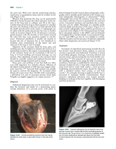
Figure 4.54. Contrast radiography can be helpful to document
synovial involvement in horses with chronic penetrating injuries of
the frog and sole. The puncture in this horse had completely healed,
Figure 4.53. Chronic penetrating injuries to the foot may be but the horse developed an abscess just above the heel bulb.
identified by small areas of granulation tissue on the sole (white Contrast injected into the abscess communicated with the navicular
arrow). bursa.