Page 512 - Adams and Stashak's Lameness in Horses, 7th Edition
P. 512
478   Chapter 4


            junction that permits infection to invade the laminae,
            which follows the path of least resistance to break open
  VetBooks.ir  may go undiagnosed until drainage at the coronary
            and drain at the coronary band. However, the condition
            band is observed. Signs of lameness may also vary
            depending on the severity and location of the infection.

            Diagnosis

              A tentative diagnosis can often be made based on the
            history and clinical signs. If pain is localized to the foot
            but there are no obvious external abnormalities, the shoe
            should be removed and the sole explored by removing the
            exfoliating  (flaky)  sole with  a hoof knife.  Acute  sole
            bruises may not be readily apparent because the hemor-
            rhage has not migrated far enough distally. Chronic
            bruises are usually visible as a stippled reddened region.
                                                           31
            In some cases the discoloration may be bluish, especially
            if a sole abscess is developing. Sole abscesses may have a
            small defect in the sole where the abscess is trying to
            break through the sole. Alternatively, a large‐bore needle
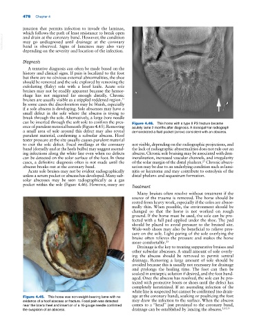
            can be inserted through the soft sole to confirm the pres-  Figure 4.46.  This horse with a type II P3 fracture became
            ence of purulent material beneath (Figure 4.45). Removing   acutely lame 2 months after diagnosis. A dorsopalmar radiograph
            a small area of sole around this defect may also reveal   demonstrated a fluid pocket (arrow) consistent with an abscess.
            purulent material, confirming a subsolar abscess. Hoof
            tester pressure at the site usually causes purulent material
            to  exit the  sole defect.  Focal swellings at the  coronary   not visible, depending on the radiographic projections, and
            band (dorsally and at the heels bulbs) may suggest ascend-  the lack of radiographic abnormalities does not rule out an
            ing infections along the white line even when no defects   abscess. Chronic sole bruising may be associated with dem-
            can be detected on the solar surface of the foot. In these   ineralization, increased vascular channels, and irregularity
            cases, a definitive diagnosis often is not made until the   of the solar margin of the distal phalanx.  Chronic absces-
                                                                                                 27
            abscess breaks out at the coronary band.           sation may be due to an underlying condition such as lami-
              Acute sole bruises may not be evident radiographically   nitis or keratoma and may contribute to osteolysis of the
            unless a serum pocket or abscess has developed. Many sub-  distal phalanx and sequestrum formation.
            solar abscesses may be seen radiographically as a gas
            pocket within the sole (Figure 4.46). However, many are   Treatment
                                                                  Many bruises often resolve without treatment if the
                                                               source of the trauma is removed. The horse should be
                                                               rested from heavy work, especially if the soles are abnor-
                                                               mally thin. When possible, the environment should be
                                                               changed so that the horse is not worked on rough
                                                               ground. If the horse must be used, the sole can be pro-
                                                               tected with a full pad applied under the shoe. The pad
                                                               should be placed to avoid pressure to the bruised site.
                                                               Wide‐web shoes may also be beneficial to relieve pres-
                                                               sure on the sole. Light paring of the sole overlying the
                                                               bruise often relieves the pressure and makes the horse
                                                               more comfortable. 25
                                                                  Drainage is the key to treating suppurative bruises and
                                                               other subsolar abscesses. A small amount of sole overly-
                                                               ing the abscess should be removed to permit ventral
                                                               drainage. Removing a large amount of sole should be
                                                               avoided because this is usually not necessary for drainage
                                                               and prolongs the healing  time.  The foot  can then be
                                                               soaked in antiseptic solution if desired, and the foot band-
                                                               aged. Once the abscess has resolved, the sole can be pro-
                                                               tected with protective boots or shoes until the defect has
                                                               completely keratinized. If an ascending infection of the
                                                               white line is suspected but cannot be confirmed (no drain-
            Figure 4.45.  This horse was non‐weight‐bearing lame with no   age at the coronary band), soaking or poulticing the foot
            evidence of a hoof abscess or fracture. Focal pain was detected   may draw the infection to the surface. When the abscess
            near the lateral heel and insertion of a 16‐gauge needle confirmed   comes to a “head” just proximal to the coronary band,
            the suspicion of an abscess.                       drainage can be established by lancing the abscess. 3,25,31

