
Page 511 - Adams and Stashak's Lameness in Horses, 7th Edition
P. 511
Lameness of the Distal Limb 477
MISCELLANEOUS CONDITIONS OF THE FOOT
VetBooks.ir gaRy M. BaxtER
SOLE BRUISES, CORNS, AND SUBSOLAR contacts the inner aspect of the shoe may also develop sole
3
ABSCESSES bruises. Any form of shoeing that concentrates weight‐
bearing on the sole is likely to cause bruising.
A bruise results from the rupture of blood vessels in Corns usually are caused by pressure from horse-
the dermis (corium or sensitive tissue) beneath the sole, shoes or when a stone becomes wedged between the
frog, or hoof wall. With time the hemorrhage spreads shoe and sole. They are rare among horses that are not
into the deep layers of the epidermis and becomes visible shod. When shoes are left on too long, the heel may
as the hoof grows. Accordingly, the discoloration associ- overgrow the shoe, causing selective pressure on the sole
ated with a sole bruise is most often seen several weeks at the angle of the wall and the bar leading to a corn.
after injury, whereas the same injury occurring in the Additionally, bending the inside branch of the shoe
hoof wall may take months before it becomes appar- toward the frog to prevent pulling or stepping off the
ent. Logically, bruises are most visible when the hem- shoe can result in direct pressure to the sole, leading to
27
orrhage is superficial and the hoof is nonpigmented. It is bruising. The application of a shoe that is one‐half to
31
likely the pain associated with the injury is due to the one full size too small for the foot also increases the
inflammatory response as well as the increased subsolar pressure on the sole area at the angle of the heels. Heel
31
pressure. If the bruised site becomes infected, a subso- calks usually enhance this effect.
31
lar abscess is likely to develop. Abscesses within the foot can develop from a variety
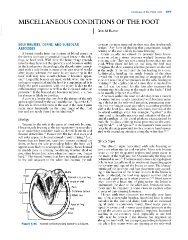
A corn is a bruise that involves the tissues of the sole of causes but are usually associated with bacteria enter-
at the angle formed by the wall and the bar (Figure 4.44). ing a defect in the sole–wall junction, penetrating inju-
31
This site is often referred to as the seat of the corn. Corns ries into the foot, or occur secondary to another problem
occur most frequently on the inner angle of the front within the hoof (i.e. laminitis, keratoma, necrosis of the
feet and are rarely found in the hindfeet. 31 collateral cartilage, etc.). For example, “quittor” is the
term used to describe necrosis and infection of the col-
Etiology lateral cartilage of the distal phalanx characterized by
multiple fistulous draining tracts proximal to the coro-
Trauma to the sole is the cause of most sole bruising. nary band, 15,31 and “gravel” is the layman’s term used to
However, sole bruising at the toe region may be secondary describe drainage proximal to the coronary band associ-
to an underlying condition such as chronic laminitis and ated with ascending infection along the white line. 22,31
flexural deformities. Horses with flat feet, thin soles, and
27
soft soles appear to be predisposed to sole bruising. Also,
9
horses that are barefoot, have their hooves trimmed too Clinical Signs
short, or have the sole protruding below the hoof wall
appear more likely to develop sole bruising. Horses housed The clinical signs associated with sole bruising or
in muddy pens in freezing conditions, whether shod or corns are often similar and variable. Most sole bruises
not, often bruise their soles when the lumpy mud freezes occur at the toe or quarter regions and corns occur at
hard. Flat‐footed horses that have repeated concussion the angle of the wall and bar. Occasionally the frog can
31
25
to the sole adjacent to the white line because the sole be bruised as well. The horse may show varying degrees
of lameness (usually mild to moderate) depending upon
the severity and type of the bruise or corn. The charac-
teristics of the lameness and foot placement vary accord-
Bulbs ing to the location of the bruise or corn. If the bruise is
Heel acute or infected, the hoof may appear warmer and an
Central sulcus of frog increased digital pulse is often present. 25,31 Hoof testers
Angle of wall often identify a focal site of pain unless the lesion is
underneath the shoe at the white line. Perineural anes-
Bars thesia may be required in some cases to exclude other
Collateral sulcus sources of pain causing lameness.
Quarter
White line Horses with foot abscesses are typically very lame
and often non‐weight bearing. Increased heat is often
palpable in the foot and distal limb and an increased
Apex of frog digital pulse is commonly found. Hoof tester pain is
typically severe, and in some cases digital pressure at the
Wall
Toe site of the abscess causes a painful response. Increased
Sole swelling at the coronary band (especially at one heel
bulb) may be present if the abscess has migrated up
along the hoof wall. For example, ascending infection of
Figure 4.44. Normal forefoot showing anatomic structures. the white line occurs when an opening at the sole–wall