
Page 506 - Adams and Stashak's Lameness in Horses, 7th Edition
P. 506
472 Chapter 4
fractures. Damage to the cartilage and subchondral
48
bone is thought to permit passage of joint fluid through
VetBooks.ir bone. 4,60 Alternatively, a developmental osteochondrosis
the opening, resulting in resorption of the subchondral
lesion predisposes to the lesion because SCLs are often
4,5
found in young horses and are frequently bilateral. See
Chapter 10 for more information about SCLs and
osteochondrosis.
Clinical Signs
A history of an acute onset of lameness may be pre-
sent, but more often the lameness is chronic and inter-
mittent. The lameness may subside with rest and recur
with exercise, and the severity can be variable. There are
often no palpable abnormalities (including hoof tester
examination), but effusion within the DIP joint may be
present. The digital pulse rate may be elevated and the A
phalangeal flexion test is usually positive. A PD nerve
27
block improves the lameness in most cases. Intrasynovial
anesthesia of the DIP joint also eliminates the lameness,
especially if the SCL communicates with the joint. 28,60
Diagnosis
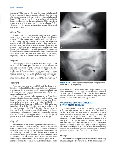
Radiography is necessary for a definitive diagnosis of
an SCL of the distal phalanx. The SCLs are variable in
size and are usually identified within the body of the dis-
tal phalanx (Figure 4.40). The majority of SCLs commu-
nicate with the joint. 28,59 In one study, 18 of 27 SCLs were
located centrally in the distal phalanx, and communica-
tion with the DIP joint was observed in all cases. In a
27
more recent study, all 11 horses had SCLs that were IA. 59
B
Treatment Figure 4.40. Lateral (A) and dorsopalmar (B) radiographs of a
horse with SCL of P3 (arrows).
Recommended treatments of SCLs of the distal pha-
lanx have included (1) confinement followed by increas-
ing exercise, (2) IA medications, (3) transcortical drilling, treated between 16 and 33 months of age, so results may
59
(4) extra‐articular surgical curettage, and (5) arthro- vary depending on the age at diagnosis. However,
scopic debridement. 28,58–60 arthroscopic debridement of SCLs of the distal phalanx
Most horses respond only transiently to IA medica- should provide a superior outcome in any age horse
tions, and debridement through hoof wall windows has compared with extra‐articular approaches.
been complicated by recurrent abscessation and lame-
ness. A dorsal arthroscopic approach for IA debridement COLLATERAL LIGAMENT INJURIES
recently has been described in 11 horses. This technique OF THE DISTAL PHALANX
59
is performed with the DIP joint extended and distracted
to permit access to the SCL. Because of the morbidity Desmitis of the CLs of the DIP joint is one of several
59
associated with extra‐articular debridement techniques, soft tissue injuries that can occur within the foot. Lesions
arthroscopic debridement should be considered the treat- of the CL of the DIP joint were the second most com-
ment of choice. 38,59 However, some SCLs in the distal mon soft tissue injury in one MRI study, and they can
phalanx may be inaccessible with the arthroscope. 28 occur alone or together with other injuries. 18–21 The
medial CL of the forelimb is the most common site of
Prognosis injury, and although uncommon, they can also occur in
the hindlimb. 19,26 These lesions may have concurrent
Variable results have been reported with extra‐articu- osseous damage to the distal phalanx at the ligament
lar debridement of these lesions, and these techniques are insertion site (Figure 4.41). 12,13 Horses with extensively
28
often complicated by infection and continued lameness. ossified collateral cartilages are also thought to be more
However, there are reports of successful debridement of prone to injuries of the CLs of the DIP joint. 21
60
SCLs through the hoof, and 1 horse treated with an
autogenous bone graft returned to race training after
58
6 months. Ten of eleven (91%) horses treated with Etiology
arthroscopic debridement of a distal phalanx SCL Asymmetrical foot placement or foot imbalances may
59
returned to athletic soundness. All of these horses were cause sliding and rotation of the distal phalanx relative