
Page 985 - Adams and Stashak's Lameness in Horses, 7th Edition
P. 985
Occupational‐Related Lameness Conditions 951
horses and potential regulatory hurdles if the horse is treated with plain hot water soaks and a softening hoof
scheduled to compete. Caution is imperative in the race pack (Animalintex) until the horn is pliant enough to
VetBooks.ir fracture exists. The results of local anesthesia are some and carry a good prognosis if managed with proper
allow eruption of the abscess. Most resolve uneventfully
horse if the possibility of anesthetizing a potential
hygiene and rest.
times open to interpretation, due to the ineffectiveness
of the block and the time frame required for anesthesia Quarter cracks occur less commonly but often enough
to develop. to be a particular nuisance. They almost always occur in
For example, a horse with proximal suspensory horses that have shearing of the heels, thin walls, an
desmitis will occasionally improve to diagnostic anes unbalanced foot, and often underslung heels, and males
thesia of the middle carpal joint. Results may also be seem to be more affected than females. The side with the
confused because excitement of the horse can obscure crack is usually taller than the unaffected side. An inte
the lameness independent of the regional anesthesia. gral component of management involves balancing the
Further ancillary diagnostic techniques include imag foot in addition to stabilizing the wall. Without balanc
ing modalities such as radiographs, ultrasound, nuclear ing the foot, repair of the crack is futile and it will recur.
scintigraphy, MRI, and computed tomography. It is rou A support shoe such as a Z‐bar shoe or a three‐quarter
tine for racetrack practitioners to be equipped with shoe with a Z bar is helpful at lessening the shearing
portable digital radiographic and ultrasonographic forces of the involved wall while healing occurs. Direct
5
equipment. Instant stall side diagnosis of fracture or repair of the crack by lacing and/or patching is often
other injury has become standard. necessary to stabilize the hoof wall. The crack must be
Nuclear scintigraphy has had a tremendous impact dry and free of sepsis, and if there is sensitive tissue
on understanding bone disease in the racehorse and the involved, a drain must be placed beneath the crack.
management of many of these conditions. Because it Many horses are able to continue training while under
provides a reflection of the physiologic status of tissue, going treatment for quarter cracks, while others must be
pathology may be detected and therefore addressed ear convalesced to allow hoof regrowth and conditioning.
lier than with plain radiography. For example, detection The farther palmar the crack is located on the foot, the
of stress‐related bone injuries may be observed before more difficult it is to manage.
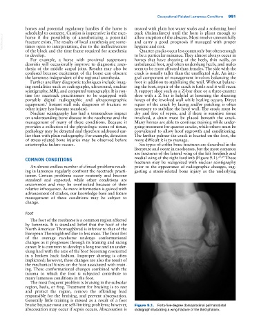
catastrophic failure occurs. Six types of coffin bone fractures are described in the
literature and occur in racehorses, but the most common
are fractures of the lateral wing of the left forelimb and
COMMON CONDITIONS medial wing of the right forelimb (Figure 9.1). 27,49 These
fractures may be recognized with nuclear scintigraphy
An almost endless number of clinical problems result prior to the appearance of radiographic changes, sug
ing in lameness regularly confront the racetrack practi gesting a stress‐related bone injury as the underlying
tioner. Certain problems occur routinely and become
standard and expected, while other conditions are
uncommon and may be overlooked because of their
relative infrequence. As more information is gained with
advancement of studies, our knowledge base and future
management of these conditions may be subject to
change.
Foot
The foot of the racehorse is a common region affected
by lameness. It is standard belief that the hoof of the
North American Thoroughbred is inferior to that of the
European Thoroughbred due to less mass. The front feet
of the average racehorse undergo conformational
changes as it progresses through its training and racing
career. It is common to develop a long toe and an under
slung heel with the axis of the foot becoming reoriented
in a broken back fashion. Improper shoeing is often
implicated; however, these changes are also the result of
the mechanical forces on the foot associated with train
ing. These conformational changes combined with the
trauma to which the foot is subjected contribute to
many lameness conditions in the foot.
The most frequent problem is bruising in the subsolar
region, heels, or frog. Treatment for bruising is to rest
and protect the region, remove the offending load
responsible for the bruising, and prevent abscessation.
Generally little training is missed as a result of a foot
bruise because most are self‐limiting problems; however, Figure 9.1. Forty‐five‐degree dorsoproximal palmarodistal
abscessation may occur if sepsis occurs. Abscessation is radiograph illustrating a wing fracture of the third phalanx.