
Page 987 - Adams and Stashak's Lameness in Horses, 7th Edition
P. 987
VetBooks.ir
A B
C
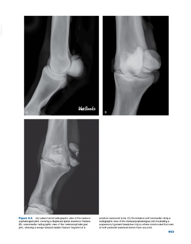
Figure 9.2. (A) Lateromedial radiographic view of the metacar- proximal sesamoid bone. (C) Dorsolateral palmaromedial oblique
pophalangeal joint, showing a displaced apical sesamoid fracture. radiographic view of the metacarpophalangeal joint illustrating a
(B) Lateromedial radiographic view of the metatarsophalangeal suspensory ligament breakdown injury where comminuted fractures
joint, showing a wedge‐shaped basilar fracture fragment of a of both proximal sesamoid bones have occurred.
953