
Page 991 - Adams and Stashak's Lameness in Horses, 7th Edition
P. 991
Occupational‐Related Lameness Conditions 957
will often reveal normal uptake, and the prognosis for
a successful career is excellent.
VetBooks.ir Humerus
Humeral fractures are a common type of injury and
are a result of an imbalance in stress remodeling.
Typically these horses demonstrate a significant lame
ness that resolves quickly with a couple days of walking.
Often a single limb is affected; however, this can be a
bilateral condition in which the horse will often demon
strate a reluctance to train and a markedly shortened
cranial phase of the stride. Clinically these horses pre
sent very similar to a scapular stress fracture; however,
stress fractures of the humerus are more common. 32,40 It
is important to note that horses with proximal suspen
sory desmitis will often demonstrate a shortened cranial
phase of their stride and diagnostic anesthesia is impor
tant in differentiating between the different regions. The
most common time to see lameness associated with the
humerus is in a horse that is returning to exercise fol
lowing a lay‐up period. It is not uncommon to see this
type of lameness while the horse is jogging or galloping
and before they complete their first breeze. The author
utilizes the humeral snap test to help diagnose horses
with humeral stress fractures. The test is performed by
holding the radius while quickly flexing the elbow joint.
A horse with a humeral stress fracture will often resent
this type of manipulation and should increase the index
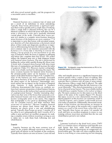
of suspicion (Video 9.1). However, nuclear scintigraphy Figure 9.8. Scintigraphic image that demonstrate an IRU on the
is the most reliable method for detecting this injury. The caudodistal aspect of the humerus.
most common location for humeral stress fractures is
the proximocaudal aspect of the humerus or caudal
aspect of the distal humerus (Figure 9.8). Complete frac older and initially present as a significant lameness that
tures of the humerus are more commonly associated resolves quickly with a couple of days of walking. This
with stress fractures of the proximocaudal aspect than is not unique to scapular stress fractures as this is a char
the distal aspect of the humerus. Interestingly race acteristic that is common with all types of stress remod
16
track surface has an impact on the location of humeral eling lameness. Often a single limb is affected. The right
stress fractures. A study performed in Southern is more commonly affected than the left; however, it can
California demonstrated that horses on synthetic sur occur bilaterally. The clinical presentation is very simi
56
faces had a greater proportion of distal humeral lesions, lar to a humeral stress fracture. The most reliable method
whereas horses on dirt surfaces had a greater proportion of identifying a scapular fracture is by nuclear scintigra
of caudoproximal lesions. Another interesting obser phy. The most common site for this injury is at the distal
16
vation was that none of the horses that sustained a com aspect of the spine of the scapula (Figure 9.9). Because
plete humeral fracture and were euthanized underwent this injury occurs in a very characteristic location, firm
nuclear scintigraphy prior to the injury. This suggests digital palpation at this location can sometimes result in
that scintigraphy is very good at identifying horses at a painful response, and a positive result would increase
risk for complete fracture and helps minimize their your index of suspicion. Additionally, ultrasound exami
occurrence in racehorses. Because complete humeral nation of this location can demonstrate an increase in
16
fractures are always fatal, horses with IRU in the thickness of the spine of the scapula with a rough bone
humerus are always given a rest and rehabilitation margin as a result of the woven bone callus that is pre
period. Typically these horses are given 3 months off if sent. Typically these horses are given 3–4 months off
no callus is seen radiographically and 4 months off if a work and have an excellent prognosis to return to race
callus is seen and have an excellent prognosis for mak training without recurrence, provided that the rest and
ing a complete return without recurrence as long as care rehabilitation period is not rushed.
is taken not to rush them back following rest and
rehabilitation.
Tarsus
Lameness localized to the distal hock joints (TMT/
Scapula DIT) is a common source of lameness and typically seen
Scapular fractures are the most common type of with horses that have sickle hock conformation.
injury to the scapula although uncommon when com Clinically these horses demonstrate an exaggerated col
pared with all injuries in the forelimb of the racehorse. lected stabby gait. The rider will often complain that the
Typically these injuries are seen in horses 3 years old and horse is cross‐firing or cross‐cantering and not changing