
Page 990 - Adams and Stashak's Lameness in Horses, 7th Edition
P. 990
956 Chapter 9
synovitis of the middle carpal joint display minimal 4+/5) initially but becomes relatively sound in a couple
lameness. The joint is often warm and there may be a of days. If the horse continues to train, it will often
VetBooks.ir normal and the condition is self‐limiting with a reduc radial carpal bones as a kissing lesion. Fragments of the
develop a fragment on the proximal intermediate and/or
change in behavior or performance. Radiographs are
distal medial radius and proximal radial carpal bone
tion in training. Joint injection is frequently performed
using a corticosteroid or IRAP in conjunction with a also commonly occur. In general, there is a good progno
chondroprotective agent. If the condition persists or sis associated with removal of fragments in the radial
recurs after treatment, a rest period of 30–60 days is carpal joint.
indicated.
Third carpal bone disease is very common in juvenile
thoroughbreds and is considered to be an integral com Radius
ponent in the development of third carpal slab or sagit Lameness associated with the radius is probably the
tal fractures. Radiographs including a skyline projection least common location in the forelimb region of the
of the third carpal bone should be performed and are Thoroughbred racehorse. However, an occasional find
diagnostic for third carpal disease. Common radio ing on nuclear scintigraphy would be an IRU in the
graphic changes are sclerosis, bone resorption beginning center of the radius classified as an enostosis‐like lesion
at the radial facet, and slab and sagittal fractures or bone islet. These findings are seen in the radius, with
1
(Figure 9.6). Prognosis is correlated with the amount the tibia and cannon bone also affected. Lameness is
21
of displacement of the fracture and degree of damage to attributed to this finding in approximately 50% of
the articular cartilage. 32 cases and correlated with the intensity of the lesion.
Other fracture configurations of the middle carpal Therefore, it is important to distinguish between a
joint include chip fracture of the distal radial carpal stress fracture and an enostosis‐like lesion, as the reha
bone, the proximal third carpal bone, and the distal bilitation program is dramatically different (Figure 9.7).
intermediate carpal bone. Prognosis for return to racing Typically, enostosis‐like lesions involving the radius
soundness depends on the extent of damage to the artic rarely result in lameness, whereas these lesions in the
ular cartilage and amount of exposed subchondral bone tibia are more likely to result in lameness. Horses with
at the load‐bearing surface. Poor prognostic signs enostosis‐like lesions are maintained in training on
include extensive cartilage damage, a void on a load‐ aspirin. These horses typically walk until they are
bearing surface, and third carpal bone involvement. Free sound and jog for 1 month before high‐intensity work
fragments in the palmar aspect of the joint are also con is resumed. A repeat nuclear scintigraphy examination
sidered a poor prognostic indicator. 24
The radial carpal joint is less frequently involved than
the middle carpal joint, but pathology affecting this
joint occurs routinely. Bone and cartilage injury usually
manifests on the distal lateral radius, which often devel
ops large (0.5–1.0 cm), partially or completely displaced
fragments. In many cases, the fragment on the distal lat
eral radius occurs first, and the horse is very lame (grade
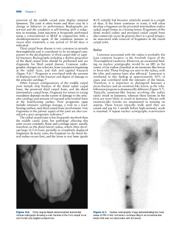
Figure 9.6. Thirty degree flexed dorsoproximal dorsodistal Figure 9.7. Nuclear scintigraphy image demonstrating two focal
oblique radiograph showing a slab fracture of the third carpal bone areas of IRU in the mid‐radius corresponding to an enostosis‐like
with frontal and sagittal components. lesion that was not associated with lameness.