
Page 992 - Adams and Stashak's Lameness in Horses, 7th Edition
P. 992
958 Chapter 9
event; however, the true etiology is unknown. Typically
this abnormality in gait is noticed when the horse is
VetBooks.ir onstrated while the horse exercises during high speeds.
walking at the barn or to the racetrack and is not dem
Oftentimes this exaggerated gait bothers the trainer
more than the horse and rarely interferes with their ath
letic potential. For these traumatic or sporadic cases, a
lateral digital extensor tenectomy utilizing a standing
approach often results in a good prognosis for improve
ment in gait; however, the exaggerated flexion of the
rear limb is often not completely eliminated.
Tibia
Tibial stress fractures are very common in 2‐year‐old
racehorses as they approach their first race. Occasionally
these fractures are seen in older horses; however, this is
much less common. Typically horses demonstrate a
hindlimb lameness following a gate work. The lameness
is most commonly identified 1–2 days following the
work, and the affected horse will demonstrate a short‐
strided, toe‐first, heel‐slapping gait when jogging in
hand in a straight line. Tibial stress fractures can occur
unilaterally or bilaterally; however, even in the bilateral
cases, one leg is often more severely affected. Nuclear
scintigraphy is the most reliable method for diagnosing
a tibial stress fracture. Typically these fractures occur
33
proximally or distally. Once a tibial stress fracture is
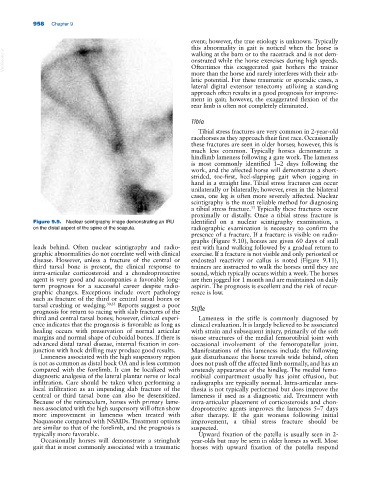
Figure 9.9. Nuclear scintigraphy image demonstrating an IRU identified on a nuclear scintigraphy examination, a
on the distal aspect of the spine of the scapula. radiographic examination is necessary to confirm the
presence of a fracture. If a fracture is visible on radio
graphs (Figure 9.10), horses are given 60 days of stall
leads behind. Often nuclear scintigraphy and radio rest with hand walking followed by a gradual return to
graphic abnormalities do not correlate well with clinical exercise. If a fracture is not visible and only periosteal or
disease. However, unless a fracture of the central or endosteal reactivity or callus is noted (Figure 9.11),
third tarsal bone is present, the clinical response to trainers are instructed to walk the horses until they are
intra‐articular corticosteroid and a chondroprotective sound, which typically occurs within a week. The horses
agent is very good and accompanies a favorable long‐ are then jogged for 1 month and are maintained on daily
term prognosis for a successful career despite radio aspirin. The prognosis is excellent and the risk of recur
graphic changes. Exceptions include overt pathology rence is low.
such as fracture of the third or central tarsal bones or
tarsal crushing or wedging. 30,55 Reports suggest a poor Stifle
prognosis for return to racing with slab fractures of the
third and central tarsal bones; however, clinical experi Lameness in the stifle is commonly diagnosed by
ence indicates that the prognosis is favorable as long as clinical evaluation. It is largely believed to be associated
healing occurs with preservation of normal articular with strain and subsequent injury, primarily of the soft
margins and normal shape of cuboidal bones. If there is tissue structures of the medial femorotibial joint with
advanced distal tarsal disease, internal fixation in con occasional involvement of the femoropatellar joint.
junction with hock drilling may produce good results. Manifestations of this lameness include the following
Lameness associated with the high suspensory region gait disturbances: the horse travels wide behind, often
is not as common as distal hock OA and is less common does not push off the affected limb normally, and has an
compared with the forelimb. It can be localized with unsteady appearance of the hindleg. The medial femo
diagnostic analgesia of the lateral plantar nerve or local rotibial compartment usually has joint effusion, but
infiltration. Care should be taken when performing a radiographs are typically normal. Intra‐articular anes
local infiltration as an impending slab fracture of the thesia is not typically performed but does improve the
central or third tarsal bone can also be desensitized. lameness if used as a diagnostic aid. Treatment with
Because of the retinaculum, horses with primary lame intra‐articular placement of corticosteroids and chon
ness associated with the high suspensory will often show droprotective agents improves the lameness 5–7 days
more improvement in lameness when treated with after therapy. If the gait worsens following initial
Naquasone compared with NSAIDs. Treatment options improvement, a tibial stress fracture should be
are similar to that of the forelimb, and the prognosis is suspected.
typically more favorable. Upward fixation of the patella is usually seen in 2‐
Occasionally horses will demonstrate a stringhalt year‐olds but may be seen in older horses as well. Most
gait that is most commonly associated with a traumatic horses with upward fixation of the patella respond