
Page 995 - Adams and Stashak's Lameness in Horses, 7th Edition
P. 995
Occupational‐Related Lameness Conditions 961
tendonitis presents as uniform enlargement of the SDFT most likely will influence the future management of ten
without fiber disruption and is often unilateral, although don injuries.
VetBooks.ir fied in 2‐year‐olds within the first several weeks or Severe injuries may require complete rest coupled with
Treatment varies according to the degree of injury.
bilateral involvement does occur. These cases are identi
systemic NSAIDs, hydrotherapy, sweating, regenerative
months when intensive training begins. As long as these
28
cases are identified prior to fiber disruption, the progno therapies, superior check ligament desmotomy, and
sis is excellent for return to training after a 60–90‐day gradual return to work, whereas minimal lesions may
rest period, and recurrence is rare. only need a reduction in training. Serial ultrasono
Physical evaluation often reveals warmth, mild to graphic examinations help determine the rate of healing
moderate edema, and pain upon palpation of an affected and ability to return to full training. Very large defects
area. Horses may exhibit palmar metacarpal swelling may benefit from surgical intervention in the form of
without SDFT swelling, usually with very low‐grade tendon splitting to permit fluid drainage from the ten
involvement of the tendon. Lameness may be nonexist don lesion.
22
ent, subtle, and transient or overt. Ultrasound examina The prognosis for return to racing is guarded follow
tion is the gold standard to gauge the tendon injury. ing tendon injuries. In general, the more severe the tear,
Caution must be used in interpretation, especially in the or the more distal the location of the lesion, the worse
acute phase, in which fluid accumulation between fibers the prognosis. Horses with superficial digital tendonitis
may resemble a tear. The follow‐up ultrasound examina in the region of the pastern have a guarded prognosis for
tion several weeks later is critical for accurate determi future racing. Tendons undergo repair in phases, and the
45
nation of the degree of tendon injury. Tears are final result is remodeled, mature scar tissue. Frequently,
recognized as hypoechoic areas within the tendon, range healed areas contain scar tissue that does not have the
dramatically in diameter and length, and may be partial elasticity of an uninjured tendon, and therefore there is
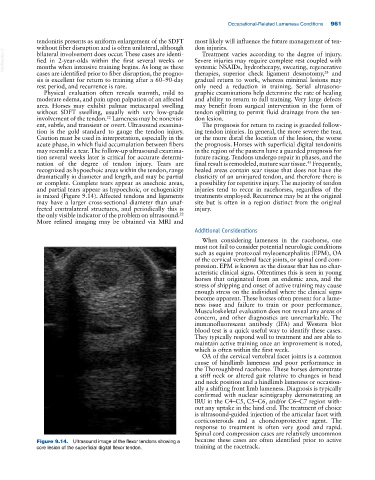
or complete. Complete tears appear as anechoic areas, a possibility for repetitive injury. The majority of tendon
and partial tears appear as hypoechoic, or echogenicity injuries tend to recur in racehorses, regardless of the
is mixed (Figure 9.14). Affected tendons and ligaments treatments employed. Recurrence may be at the original
may have a larger cross‐sectional diameter than unaf site but is often in a region distinct from the original
fected contralateral structures, and periodically this is injury.
the only visible indicator of the problem on ultrasound.
22
More refined imaging may be obtained via MRI and
Additional Considerations
When considering lameness in the racehorse, one
must not fail to consider potential neurologic conditions
such as equine protozoal myleoencephalitis (EPM), OA
of the cervical vertebral facet joints, or spinal cord com
pression. EPM is known as the disease that has no char
acteristic clinical signs. Oftentimes this is seen in young
horses that originated from an endemic area, and the
stress of shipping and onset of active training may cause
enough stress on the individual where the clinical signs
become apparent. These horses often present for a lame
ness issue and failure to train or poor performance.
Musculoskeletal evaluation does not reveal any areas of
concern, and other diagnostics are unremarkable. The
immunofluorescent antibody (IFA) and Western blot
blood test is a quick useful way to identify these cases.
They typically respond well to treatment and are able to
maintain active training once an improvement is noted,
which is often within the first week.
OA of the cervical vertebral facet joints is a common
cause of hindlimb lameness and poor performance in
the Thoroughbred racehorse. These horses demonstrate
a stiff neck or altered gait relative to changes in head
and neck position and a hindlimb lameness or occasion
ally a shifting front limb lameness. Diagnosis is typically
confirmed with nuclear scintigraphy demonstrating an
IRU in the C4–C5, C5–C6, and/or C6–C7 region with
out any uptake in the hind end. The treatment of choice
is ultrasound‐guided injection of the articular facet with
corticosteroids and a chondroprotective agent. The
response to treatment is often very good and rapid.
Spinal cord compression cases are relatively uncommon
Figure 9.14. Ultrasound image of the flexor tendons showing a because these cases are often identified prior to active
core lesion of the superficial digital flexor tendon. training at the racetrack.