
Page 994 - Adams and Stashak's Lameness in Horses, 7th Edition
P. 994
960 Chapter 9
with pelvic stress fractures demonstrate a significant horses will jog sound when examined in hand. The SI
lameness following a gate work and are often pulled up region is medicated with corticosteroids and sarapin; if
VetBooks.ir Additionally these horses demonstrate a very sore, addressed as well.
the primary lameness is identified, this region should be
by the rider shortly after breaking out of the gate.
Soft tissue injuries are recognized in the hip area and
tucked up hind end, and short‐strided gait, and the
trainer often believes the horse is tied up. Horses with are believed to occur during propulsion at fast work.
suspected pelvic fractures should be maintained tied to a Synthetic surfaces with increased traction have been
wire in the stall to keep them standing for a minimum of incriminated in development of this condition; however,
30 days. Recumbency increases the risk of complete there is no current evidence to support this theory.
fracture, which can result in laceration of closely associ Treatment consists of rest, and the prognosis is good for
ated vessels causing fatal hemorrhage. Special care full recovery.
should be taken to monitor these horses for pneumonia
as this is a complication of maintaining a horse on a tie
wire with the head elevated. The most common location Tendons and Ligaments
is the ilial wing, and nuclear scintigraphy is the most Tendon and ligament injuries in racing Thoroughbreds
reliable method of detecting this injury and its location occur predominantly from intrinsic sources, although
(Figure 9.12). Ultrasound examination is often impor extrinsic trauma or injury occasionally occurs. The etiol
tant in characterizing the type of fracture. Prognosis for ogy is multifactorial, but cumulative excessive strain
most injuries in this region is favorable with rest as long rates in addition to the gradual demise of structural sta
as the fracture is not displaced. Typically horses with bility incurred with degenerative aging changes that are
ileal wing fractures are given 3–4 months off and have accelerated by exercise are the primary components.
52
an excellent prognosis with minimal risk of recurrence, Repetitive loading of a compromised tendon (one that
whereas fractures that are comminuted, articular, or has sustained subclinical, microscopic damage) may be
involve the acetabulum have a poor prognosis for return responsible for the often progressive nature of these
to racing, and an alternative career should be injuries. Incidence of tendon injury is high and in one
47
considered. study was reported to be responsible for up to 46% of
Less commonly, lameness will be localized to the SI all racetrack injuries, although in clinical experience the
2
joints. SI pain is often secondary to a primary lameness rate is not this high.
lower in the limb, resulting in a change in gait and the The forelimb superficial digital flexor tendon (SDFT)
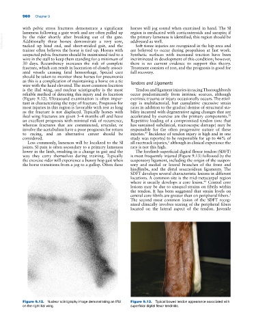
way they carry themselves during training. Typically is most frequently injured (Figure 9.13) followed by the
the exercise rider will experience a bunny hop gait when suspensory ligament, including the origin of the suspen
the horse transitions from a jog to a gallop. Often these sory and medial or lateral branches of the front and
hindlimbs, and the distal sesamoidean ligaments. The
SDFT develops several characteristic lesions in different
locations. A common site is the mid‐metacarpal region
45
where it usually develops a core lesion. Central core
lesions may be due to unequal strains on fibrils within
the tendon. It has been suggested that strain levels on
central core fibrils are greater than on peripheral fibers.
3
The second most common lesion of the SDFT recog
nized clinically involves tearing of the peripheral fibers
located on the lateral aspect of the tendon. Juvenile
Figure 9.12. Nuclear scintigraphy image demonstrating an IRU Figure 9.13. Typical bowed tendon appearance associated with
on the right ilial wing. superficial digital flexor tendinitis.