
Page 988 - Adams and Stashak's Lameness in Horses, 7th Edition
P. 988
954 Chapter 9
of abaxial fragments is routinely performed. Generally,
if marked displacement has occurred, there are usually
VetBooks.ir return to racing. However, if there is minimal suspen
excessive suspensory damage and a poor prognosis for
sory damage, surgical removal can have a favorable out
come to return to race, but only a fair prognosis for
returning at the same level. In general, horses with frac
tures of the lateral sesamoid bone do better than those
with fractures involving the medial sesamoid bone, and
hindlimbs do better than forelimbs. Midbody fractures
may be managed conservatively or with surgical treat
25
ment with lag screw fixation or circumferential wiring.
Contrary to literature reports, there is a guarded prog
nosis for future racing. One report that supports this
demonstrated a 44% return to racing when lag screw
fixation was elected and 0% when circumferential wir
ing was elected. Radiographs often indicated that the
8
fractures have completely healed; however, once back in
training, lameness often occurred when the horse
worked 5/8th of a mile and re‐injury was common.
Degenerative joint changes often accompanied these
injuries and worsened the prognosis. With conservative
management, horses usually become sound and are use
ful for breeding or light athletic use.
Stress‐related bone disease of the distal metacar
pus or metatarsus is an extremely common cause of
lameness in juvenile horses and is also seen in older
horses. The most common clinical manifestation is a
variable severity of lameness, usually seen when cool
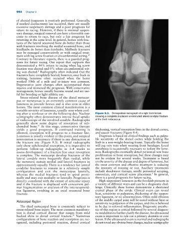
ing after a workout or on the following day. Nuclear Figure 9.3. Dorsopalmar radiograph of a right front fetlock
scintigraphy often demonstrates intense focal uptake showing a complete displaced comminuted lateral condylar fracture
of radioisotope of the involved condyle. Radiographs of the third metacarpus.
generally show some degree of resorption of sub
chondral bone. At this stage, conservative therapy
31
yields a good prognosis. If continued training is thickening, vertical resorption lines in the dorsal cortex,
allowed, resorption will progress to a fracture line. and saucer fractures (Figure 9.4).
Lameness is usually evident at this point, and surgical Diagnosis is based on clinical findings such as palpa
intervention is necessary if there is any distraction at ble sensitivity of the dorsal cortex while holding the
the fracture site (Figure 9.3). If initial radiographs limb in a non‐weight‐bearing frame. Typically the horse
only show subchondral resorption, it is imperative to will jog very sore when wearing front bandages. Local
perform follow‐up radiographs in 3–4 weeks to anesthesia is occasionally necessary to isolate the lame
assess development of a fracture line once resorption ness. Radiographs eventually detect periosteal new bone
is complete. The metacarpi develop fractures of the proliferation or bone resorption, but these changes may
lateral condyle more frequently than medial, while not be evident for several weeks. Treatment is based
the metatarsi sustain medial and lateral fractures in on the severity of the disease and degree of lameness, but
approximately equally. There is a tendency for lateral the most common and effective treatment is reducing
condylar fractures to remain in a relatively simple the intensity of training or rest. Ancillary treatments
configuration and exit the metacarpus laterally, include shockwave therapy, needle periosteal scraping,
9
whereas the medial fractures tend to spiral proxi osteostixis, and cortical screw placement. In general,
mally and may exit medially or laterally. Prognosis is there is a good prognosis for future racing.
directly correlated with the extent of articular injury High suspensory lameness in the forelimb presents in
and if the fracture is displaced with concurrent pal a variety of different ways and can be a diagnostic chal
mar fragmentation or avulsion of the intersesamoid lenge. Clinically these horses demonstrate a shortened
ean ligament, resulting in an axial sesamoid bone cranial phase of the stride. Clinical exam can reveal
fracture. heat, sensitivity to palpation, thickening of the suspen
sory ligament, or no abnormalities. Often mild effusion
Metacarpal Region of the middle carpal joint will be noted without heat or
sensitivity to palpation of the carpus, and this is believed
The third metacarpal bone is commonly subject to to be due to referred inflammation. Diagnostic anesthe
stress‐related bone injury. The most common manifesta sia of this region is critical followed by several diagnos
tion is dorsal cortical disease that ranges from mild tic modalities to further clarify the disease. An ultrasound
bucked shins to dorsal cortical fracture. Numerous exam is important to rule out a primary desmitis or core
39
configurations of bone reaction and resorption are rec lesion. If the ultrasound exam is normal and radiographs
ognized, including periosteal reaction, dorsal cortical do not reveal any obvious bony changes, nuclear scintigraphy