
Page 257 - Anatomy and Physiology of Farm Animals, 8th Edition
P. 257
242 / Anatomy and Physiology of Farm Animals
The utriculus and the smaller sacculus
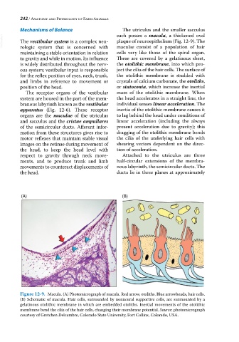
Mechanisms of Balance each posses a macula, a thickened oval
VetBooks.ir The vestibular system is a complex neu plaque of neuroepithelium (Fig. 12‐9). The
maculae consist of a population of hair
rologic system that is concerned with
maintaining a stable orientation in relation cells very like those of the spiral organ.
to gravity and while in motion. Its influence These are covered by a gelatinous sheet,
is widely distributed throughout the nerv the otolithic membrane, into which pro
ous system; vestibular input is responsible ject the cilia of the hair cells. The surface of
for the reflex position of eyes, neck, trunk, the otolithic membrane is studded with
and limbs in reference to movement or crystals of calcium carbonate, the otoliths,
position of the head. or statoconia, which increase the inertial
The receptor organs of the vestibular mass of the otolithic membrane. When
system are housed in the part of the mem the head accelerates in a straight line, the
branous labyrinth known as the vestibular individual senses linear acceleration. The
apparatus (Fig. 12‐6). These receptor inertia of the otolithic membrane causes it
organs are the maculae of the utriculus to lag behind the head under conditions of
and sacculus and the cristae ampullares linear acceleration (including the always
of the semicircular ducts. Afferent infor present acceleration due to gravity); this
mation from these structures gives rise to dragging of the otolithic membrane bends
motor reflexes that maintain stable visual the cilia of the underlying hair cells with
images on the retinae during movement of shearing vectors dependent on the direc
the head, to keep the head level with tion of acceleration.
respect to gravity through neck move Attached to the utriculus are three
ments, and to produce trunk and limb half‐circular extensions of the membra
movements to counteract displacements of nous labyrinth, the semicircular ducts. The
the head. ducts lie in three planes at approximately
(A) (B)
Figure 12-9. Macula. (A) Photomicrograph of macula. Red arrow, otoliths. Blue arrowheads, hair cells.
(B) Schematic of macula. Hair cells, surrounded by nonneural supportive cells, are surmounted by a
gelatinous otolithic membrane in which are embedded otoliths. Inertial movements of the otolithic
membrane bend the cilia of the hair cells, changing their membrane potential. Source: photomicrograph
courtesy of Gretchen Delcambre, Colorado State University, Fort Collins, Colorado, USA.