
Page 444 - Anatomy and Physiology of Farm Animals, 8th Edition
P. 444
The Urinary System / 429
cells contain the enzyme renin. Renin is a fenestrated (i.e., has openings or pores in
the cells), so this part of the barrier is highly
component of the renin–angiotensin–
VetBooks.ir aldosterone system (see Chapter 18, permeable. Podocytes (cells of the inner
layer of the glomerular capsule) have cellu-
Fig. 18‐11), which is involved in the regula-
tion of blood volume and blood pressure. lar extensions that rest on the glomerular
The JG cells are part of a functional basement membrane, but slit‐like pores
grouping of closely related structures, between the extensions permit the passage
the juxtaglomerular apparatus. The of the filtrate (Fig. 23‐8).
juxtaglomerular apparatus consists of the The glomerular filtration barrier acts
JG cells, the macula densa, and extraglo- much like a sieve, and all substances up to
merular mesangial cells (Fig. 23‐7). The a molecular weight of about 65,000 pass
macula densa is a specific region of the through the barrier. Blood cells are too
wall of the distal tubule where the cellular large to pass, and only a small percentage
nuclei appear to be bunched closely of plasma proteins pass through the bar-
together. The segment of the distal tubule rier. Most other plasma constituents (e.g.,
found here is part of the same nephron glucose, amino acids, urea, creatinine,
associated with the afferent arterioles sodium, potassium, chlorine, and bicarbo-
(Fig. 23‐3). The extraglomerular mesangial nate ions) readily cross the barrier, and their
cells are between the macula densa and its concentrations in the initial filtrate are
associated JG cells. about the same as in plasma. Proteinuria is
the presence of abnormal amounts of
protein in voided urine. Kidney diseases
Glomerular Filtration that localize in or primarily affect
glomeruli are often associated with
The glomerular filtrate is the fluid and fluid proteinuria or hematuria (blood in
constituents that pass from the blood voided urine).
plasma in the glomerulus into the urinary The forces determining the rate of
space of the glomerular capsule. The physi- movement of fluid across the glomerular
cal barriers through which the filtrate passes filtration barrier are the same as those that
include: (1) the capillary endothelium of the determine fluid movement out of capillar-
glomerulus; (2) the inner layer of the ies throughout the body. The effective
glomerular capsule; and (3) a basement filtration pressure (the pressure tending
membrane (lamina) between these two cell to force fluid out of the capillary) is usually
layers. The glomerular endothelium is considered to be the difference between
p
us
p
pp
e
ep
fs
e fp gbm ep
p
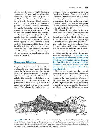
Figure 23-8. The glomerular filtration barrier. e, fenestrated glomerular capillary endothelium; ep,
endothelial pore (fenestra); gbm, glomerular basement membrane; p, podocytes, or visceral layer of
Bowman’s capsule with their filtration slits (FS); us, urinary space of Bowman’s capsule. Source: Dellmann
and Eurell, 1998. Reproduced with permission of John Wiley & Sons, Inc.