
Page 440 - Anatomy and Physiology of Farm Animals, 8th Edition
P. 440
The Urinary System / 425
VetBooks.ir Arcuate artery Interlobular artery
Afferent
Efferent arteriole
Nephron loop arteriole
Glomerulus
Glomerular
capsule
Vasa recta
Proximal
tubule
Distal tubule
Collecting duct
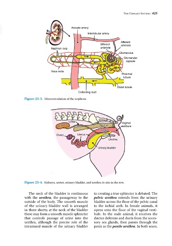
Figure 23-3. Microcirculation of the nephron.
Ureter Vaginal
Kidney Rectum vestibule
Vagina
Ovary Uterus
Urethra
Urinary bladder
Figure 23-4. Kidneys, ureter, urinary bladder, and urethra in situ in the sow.
The neck of the bladder is continuous in creating a true sphincter is debated. The
with the urethra, the passageway to the pelvic urethra extends from the urinary
outside of the body. The smooth muscle bladder across the floor of the pelvic canal
of the urinary bladder wall is arranged to the ischial arch. In female animals, it
in three sheets; at the neck of the bladder opens onto the floor of the vaginal vesti-
these may form a smooth muscle sphincter bule. In the male animal, it receives the
that controls passage of urine into the ductus deferens and ducts from the acces-
urethra, although the precise role of the sory sex glands, then passes through the
intramural muscle of the urinary bladder penis as the penile urethra. In both sexes,