
Page 439 - Anatomy and Physiology of Farm Animals, 8th Edition
P. 439
424 / Anatomy and Physiology of Farm Animals
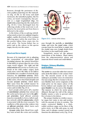
however, through the persistence of dis- Arcuate
crete papillae projecting into the renal pel-
VetBooks.ir vis (Fig. 23‐1B). In the horse and small Interlobar arteries Glomerulus
arteries
ruminants, the individual papillae, like the
cortex, are fused. Consequently, they pre-
sent as a single longitudinal ridge, the
renal crest, projecting into the renal pelvis
(Fig. 23‐1C). Urine discharged from the
collecting tubules of the renal crest is col- Renal Interlobular
lected in the renal pelvis and from there is artery artery
delivered to the ureter.
In the kidney of the ox and pig, individ-
ual pyramids project into calyces (singular,
calyx), cuplike diverticula of the common
collecting space within the renal hilus. In Figure 23-2. Arteries of the kidney.
the porcine kidney these empty into the
renal pelvis. The bovine kidney has no pass through the medulla as interlobar
pelvis and so the calyces in this species veins, and enter the renal veins, which
empty directly into the ureter. emerge from the renal hilus to empty into
the caudal vena cava. Lymph drains from
the kidney to the renal lymph nodes.
Blood and Nerve Supply Sympathetic nerves are the primary
innervation of the kidneys. These derive
Because of its important role in adjusting from the celiacomesenteric plexus and
the composition of extracellular fluid innervate blood vessels and renal tubules.
(including plasma), the arterial blood flow
to the kidney is greater than the size of the
organ would suggest. The right and left Ureters, Urinary Bladder,
renal arteries may receive as much as one‐ and Urethra
fourth of the total cardiac output. Each
renal artery enters the hilus of the kidney The ureter is a muscular tube that conveys
and divides into a number of relatively large urine from the kidney to the urinary blad-
branches, the interlobar arteries. These der. The smooth muscle of the ureter
pass peripherally between pyramids almost undergoes peristaltic waves of contraction
to the cortex, where they bend abruptly and that encourage the flow of urine to the uri-
become arcuate arteries, which derive nary bladder. Each ureter originates at the
their name from the arched manner by renal pelvis (or the major calices of the
which they pass along the junction between bovine kidney) and empties into the uri-
cortex and medulla (Fig. 23‐2). nary bladder near its neck at the trigone.
Each arcuate artery gives off a number The manner in which the ureter passes
of interlobular arteries that extend into obliquely through the wall of the urinary
the cortex and in turn give rise to the affer- bladder creates a valve to prevent reflux
ent arterioles. Each afferent arteriole flow of urine to the kidney.
branches repeatedly to form a tufted capil- The urinary bladder is a hollow muscu-
lary network called the glomerulus, which lar organ that varies in size and position
is associated with the renal corpuscle. The with the amount of urine it contains. The
capillaries of the glomerulus coalesce into empty, contracted bladder is a thick‐
an efferent arteriole, which leaves each walled, piriform organ on the floor of the
glomerulus (Fig. 23‐3). pelvic cavity. As it fills with urine, its wall
Arcuate veins drain blood from capil- thins, and it enlarges craniad toward and
lary beds in both the cortex and medulla, then into the abdominal cavity (Fig. 23‐4).