
Page 181 - Clinical Pearls in Cardiology
P. 181
Ischemic Heart Disease 169
A total score of 3 has a high specificity for diagnosing
acute myocardial infarction in a person with chest pain
who has LBBB pattern in his ECG if no previous ECG
tracings are available with him or in a person with chest
pain who has preexisting LBBB pattern in his previous
electrocardiograms. Cabrera’s sign is a notching in the
ascending limb of the S wave in lead V3 or V4. Chapman’s
sign is a notching in the ascending limb of the R wave
in lead I, aVL or V6. Both are features of old myocardial
infarction in the presence of LBBB pattern.
21. What are the characteristics of ischemic T wave
inversion?
T wave inversion is a very nonspecific finding. Inverted T
waves are normal in leads III, aVR and V1 in association
with a predominantly negative QRS complex. T wave
inversion in lead III is reversed on deep inspiration.
Many conditions like bundle branch block, ventricular
hypertrophy with strain, etc. are also associated with
T wave inversion. The clues that help to distinguish
ischemic T wave inversion from that due to other causes
are the following:
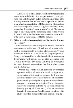
• T waves due to myocardial ischemia are inverted
symmetrically and have a pointed configuration,
whereas in most other circumstances the T waves are
asymmetrically inverted (“reverse checkmark”
pattern with gradually slanting downslope and steep
upslope). However, symmetrical T wave inversion
can occur normally in leads V1–V3 of an otherwise
healthy young adult woman (called as persistent
juvenile T wave pattern) and in some conditions like
HCM (called as pseudoinfarction pattern) (Fig. 8).