
Page 184 - Clinical Pearls in Cardiology
P. 184
172 Clinical Pearls in Cardiology
then the same leads would show ST depression in a
posterior wall myocardial infarction. This characteristic
electrocardiographic effect is called as ‘reciprocity’.
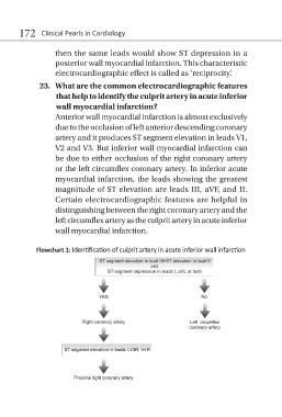
23. What are the common electrocardiographic features
that help to identify the culprit artery in acute inferior
wall myocardial infarction?
Anterior wall myocardial infarction is almost exclusively
due to the occlusion of left anterior descending coronary
artery and it produces ST segment elevation in leads V1,
V2 and V3. But inferior wall myocardial infarction can
be due to either occlusion of the right coronary artery
or the left circumflex coronary artery. In inferior acute
myocardial infarction, the leads showing the greatest
magnitude of ST elevation are leads III, aVF, and II.
Certain electrocardiographic features are helpful in
distinguishing between the right coronary artery and the
left circumflex artery as the culprit artery in acute inferior
wall myocardial infarction.
Flowchart 1: Identification of culprit artery in acute inferior wall infarction