
Page 183 - Clinical Pearls in Cardiology
P. 183
Ischemic Heart Disease 171
22. How do you localize ischemic myocardial territory
from the electrocardiogram?
The various electrocardiographic leads look at the
heart from different angles. So changes to the pattern
of elecrocardiographic recording due to myocardial
ischemia manifest in different ways in these leads,
depending upon the location of ischemic territory.
The inferior leads (II, III and aVF) look at the right
and diaphragmatic side of the heart and this territory is
perfused mainly by the right coronary artery (RCA). So
ischemia of this part of the heart produces changes in
the II, III and aVF leads. The anterior leads (V2, V3 and
V4) look at the front or anterior part of the heart, which
is comprised mainly by the left ventricle. This territory is
supplied by the left anterior descending artery (LAD). So
ischemia of this region produces changes in the V2, V3
and V4 leads. The lateral leads (I, aVL, V5 and V6) look
at the left side of the heart and they reflect the lateral
part of the left ventricle. This part of the heart is perfused
by the left circumflex artery (LCX). So ischemia of this
region produces changes in the I, aVL, V5 and V6 leads.
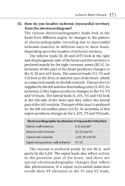
Electrocardiographic localization of myocardial infarction
Inferior wall ischemia II, III and aVF
Anterior wall ischemia V2, V3 and V4
Lateral wall ischemia I, aVL, V5 and V6
Septal and posterior wall ischemia V1, V2
The septum is perfused partly by the RCA, and
partly by the LAD. The septal leads also reflect activity
in the posterior part of the heart, and there are
special electrocardiographic changes that reflect
this phenomenon. If a septal myocardial infarction
would show ST elevation in the V1 and V2 leads,