
Page 185 - Clinical Pearls in Cardiology
P. 185
Ischemic Heart Disease 173
ECG confirmation of the infarct-related artery during
acute inferior myocardial infarction may be particularly
valuable when coronary angiography indicates lesions
in both the right and left circumflex coronary arteries.
In patients with acute inferior myocardial infarction, ST
depression in leads V1–V3 has been shown by numerous
investigators to indicate a larger infarction with extension
of the injury to the posterolateral and/or the inferoseptal
wall. Such ST depression in these ‘anterior’ leads during
acute inferior wall MI is a reciprocal change and does not
indicate concomitant LAD occlusive disease. It is seen
in both right coronary artery and left circumflex related
inferior infarctions.
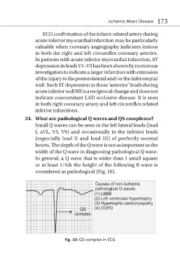
24. What are pathological Q waves and QS complexes?
Small Q waves can be seen in the left lateral leads (lead
I, aVL, V5, V6) and occasionally in the inferior leads
(especially lead II and lead III) of perfectly normal
hearts. The depth of the Q wave is not as important as the
width of the Q wave in diagnosing pathological Q wave.
In general, a Q wave that is wider than 1 small square
or at least 1/4th the height of the following R wave is
considered as pathological (Fig. 10).
Fig. 10: QS complex in ECG