
Page 158 - Essential Haematology
P. 158
144 / Chapter 10 Spleen
severe haemolytic and megaloblastic anaemias.
Extramedullary haemopoiesis may result either
from reactivation of dormant stem cells within the
spleen or homing of stem cells from the bone
marrow to the spleen.
Imaging the s pleen
Ultrasound is the most frequently used technique
to image the spleen (Fig. 10.3 ). This can also detect
whether or not blood flow in the splenic, portal and
hepatic veins is normal, as well as liver size and
consistency. Computed tomography (CT) is prefer-
able for detecting structural detail and any associ-
ated lymphadenopathy (e.g. for lymphoma staging).
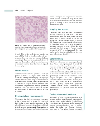
Figure 10.2 Splenic atrophy: peripheral blood fi lm
Magnetic resonance imaging (MRI) also gives
showing Howell – Jolly bodies, Pappenheimer bodies
improved fine detail structure. Positron emission
(siderotic granules; see p. 30 ) and misshapen cells.
tomography (PET) is used particularly for initial
staging and for detecting residual disease after treat-
(Howell – Jolly bodies) and siderotic granules are ment of lymphoma (Fig. 10.4 ).
removed (Fig. 10.2 ). In the relatively hypoxic envi-
ronment of the red pulp, and because of plasma Splenomegaly
skimming in the cords, the membrane fl exibility of
aged and abnormal red cells is impaired and they Splenic size is increased in a wide range of condi-
are retained within the sinus where they are ingested tions (Table 10.1 ). Splenomegaly is usually felt
by macrophages. under the left costal margin but massive splenom-
egaly may be felt in the right iliac fossa (see Fig.
15.4 ). The spleen moves with respiration and a
Immune f unction
medial splenic notch may be palpable in some cases.
The lymphoid tissue in the spleen is in a unique In developed countries the most common causes of
position to respond to antigens filtered from the splenomegaly are infectious mononucleosis, haema-
blood and entering the white pulp. Macrophages tological malignancy and portal hypertension,
and dendritic cells in the marginal zone initiate an whereas malaria and schistosomiasis are more preva-
immune response and then present antigen to B and lent on a global scale (Table 10.1 ). Chronic myeloid
T cells to start adaptive immune responses. Th is leukaemia, primary myelofi brosis, lymphoma,
arrangement is highly efficient at initiating immune Gaucher ’ s disease, malaria, leishmaniasis and
responses to encapsulated bacteria and explains schistosomiasis are potential causes of massive
the susceptibility of hyposplenic patients to these splenomegaly.
organisms.
Tropical s plenomegaly s yndrome
Extramedullary h aemopoiesis
A syndrome of massive splenomegaly of uncertain
The spleen, like the liver, undergoes a transient aetiology has been found frequently in many malar-
period of haemopoiesis at around 3 – 7 months of ious zones of the tropics including Uganda, Nigeria,
fetal life but is not a site of erythropoiesis in the New Guinea and the Congo. Smaller numbers of
adult. However, haemopoiesis may be re - established patients with this disorder are seen in southern
in both organs as extramedually haemopoiesis , in Arabia, the Sudan and Zambia. Previously,
disorders such as primary myelofibrosis or in chronic such terms as ‘ big spleen disease ’ , ‘ cryptogenic