
Page 160 - Essential Haematology
P. 160
146 / Chapter 10 Spleen
(a) (b)
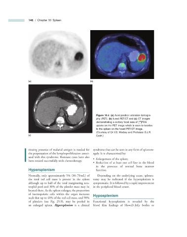
Figure 10.4 (a) Axial positron emission tomogra-
phy (PET), (b) fused PET/CT and (c) CT images
18
demonstrating a solitary focal area of [ ]FDG
uptake on the PET image which is seen to localize
to the spleen on the fused PET/CT image.
(Courtesy of Dr V.S. Warbey and Professor G.J.R.
(c) Cook .)
tinuing presence of malarial antigen is needed for syndrome that can be seen in any form of splenom-
the perpetuation of the lymphoproliferation associ- egaly. It is characterized by:
ated with this syndrome. Resistant cases have also
• Enlargement of the spleen;
been treated successfully with chemotherapy.
• Reduction of at least one cell line in the blood
in the presence of normal bone marrow
Hypersplenism function.
Normally, only approximately 5% (30 – 70 mL) of Depending on the underlying cause, splenec-
the total red cell mass is present in the spleen tomy may be indicated if the hypersplenism is
although up to half of the total marginating neu- symptomatic. It is followed by a rapid improvement
trophil pool and 30% of the platelet mass may be in the peripheral blood count.
located there. As the spleen enlarges, the proportion
of haemopoietic cells within the organ increases Hyposplenism
such that up to 40% of the red cell mass, and 90%
of platelets (see Fig. 25.9 ), may be pooled in Functional hyposplenism is revealed by the
an enlarged spleen. Hypersplenism is a clinical blood fi lm findings of Howell – Jolly bodies or