
Page 186 - Essential Haematology
P. 186
172 / Chapter 12 Haematological malignancy: management
(a) (b)
(c) (d)
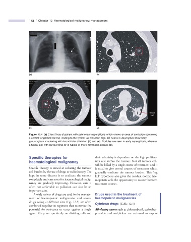
Figure 12.4 (a) Chest X - ray of patient with pulmonary aspergillosis which shows an area of cavitation containing
a central fungal ball (arrow) leading to the typical ‘ air - crescent ’ sign. CT scans in Aspergillus show hazy
ground - glass shadowing with bronchiolar dilatation (b) and (c) . Nodules are seen in early aspergillosis, whereas
a fungal ball with surrounding air is typical of more advanced disease (d) .
Specifi c t herapies for their selectivity is dependent on the high prolifera-
h aematological m alignancy tion rate within the tumour. Not all tumour cells
will be killed by a single course of treatment and it
Specific therapy is aimed at reducing the tumour is usual to give several courses of treatment which
cell burden by the use of drugs or radiotherapy. Th e gradually eradicate the tumour burden. This ‘ log
hope in some diseases is to eradicate the tumour kill ’ hypothesis also gives the residual normal hae-
completely and cure rates for haematological malig- mopoietic cells the opportunity to recover between
nancy are gradually improving. However, cure is treatment courses.
often not achievable so palliation can also be an
important aim.
A wide variety of drugs are used in the manage- Drugs u sed in the t reatment of
ment of haemopoietic malignancies and several h aemopoietic m alignancies
drugs acting at different sites (Fig. 12.5 ) are often
Cytotoxic d rugs (Table 12.1 )
combined together in regimens that minimize the
potential for resistance to occur against a single Alkylating agents such as chlorambucil, cyclophos-
agent. Many act specifically on dividing cells and phamide and melphalan are activated to expose