Page 223 - Essential Haematology
P. 223
Chapter 15  Myeloproliferative neoplasms  /  209


                      tion and overproduction of platelets. Th e haemat-      Clinical and  l aboratory  fi ndings
                      ocrit is normal and the Philadelphia chromosome

                                                                 The dominant clinical features are thrombosis and
                      or  BCR - ABL1  rearrangement are absent. Th e bone
                                                                haemorrhage. Most cases are symptomless and diag-
                      marrow shows no collagen fibrosis. A persisting   nosed on routine blood counts. Th rombosis  may

                                               9
                      platelet count of  > 450    ×    10  /L is the central
                                                                occur in the venous or arterial systems (Fig.  15.5 b)
                      diagnostic feature but other causes of a raised
                                                                whereas haemorrhage, as a result of abnormal plate-
                      platelet count (particularly iron defi ciency, infl am-
                                                                let function, may cause either chronic or acute
                      matory or malignant disorder and myelodysplasia)
                                                                bleeding. Some patients (JAK2 + ) present with
                      need to be fully excluded before the diagnosis can
                                                                Budd – Chiari syndrome when the platelet count
                      be made.
                                                                may be normal because of the splenomegaly. A char-
                          Half of patients show the  JAK2   (Val617Phe)
                                                                acteristic symptom is erythromelalgia, a burning
                      mutation and these cases tend to resemble more
                                                                sensation felt in the hands or feet and promptly
                      closely PV with higher haemoglobin and white cell
                                                                relieved by aspirin. Up to 40% of patients will have
                      counts than  JAK2  negative cases. Mutations within
                                                                palpable splenomegaly whereas in others there may
                      the  MPL  gene are seen in 4% of cases. Rare primary
                                                                be splenic atrophy because of infarction.
                      familial cases in children have been associated with
                                                                     Abnormal large platelets and megakaryocyte
                      mutations in the genes for thrombopoietin or its
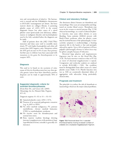
                                                                fragments may be seen on the blood fi lm  (Fig.
                      receptor MPL.
                                                                 15.8 ). The bone marrow is similar to that in PV but

                                                                an excess of abnormal megakaryocytes is typical.
                                                                Cytogenetics and molecular analysis are analysed
                          Diagnosis                             to exclude BCR - ABL1 +  CML. Th e  condition
                       This used to be based on the exclusion of other   must be distinguished from other causes of a raised

                      causes of chronic thrombocytosis but now that spe-  platelet count (Table  15.5 ). Platelet function tests

                      cific genetic lesions have been identified a positive   (see p. 328  ) are consistently abnormal, failure of

                      diagnosis can be made in approximately 50% of   aggregation with adrenaline being particularly
                      cases.                                    characteristic.
                                                                    Prognosis and  t reatment
                          Suggested  d iagnostic  c riteria for    The principle is to reduce the risk of thrombosis or

                        e ssential  t hrombocythaemia           haemorrhage which are the major clinical problems.
                      (From Beer P.A. and Green A.R. (2009)
                        Hematology Am Soc Hematol Educ Program ,
                      621 – 8.)
                        Diagnosis requires A1 – A3 or A1  +  A3 – A5:
                                                     9
                         A1      Sustained platelet count  > 450    ×    10  /L.

                         A2   Presence of an acquired pathogenetic mutation


                          (e.g. in  JAK2  or  MPL ).
                         A3   No other myeloid malignancy, PV, primary


                          myelofibrosis, chronic myeloid leukaemia
                          (CML) or myelodysplastic syndrome.
                         A4      No reactive cause for thrombocytosis and
                          normal iron stores.
                         A5      Bone  marrow  trephine  histology  showing
                          increased megakaryocytes with prominent large             Figure 15.8   Peripheral blood fi lm in essential


                          hyperlobulated forms; reticulin is generally not   thrombocythaemia showing increased numbers of
                          increased.                            platelets and a nucleated megakaryocytic fragment.

