
Page 24 - Essential Haematology
P. 24
10 / Chapter 1 Haemopoiesis
in their dimerization and translocation from the cell complex cascades of biochemical events resulting in
cytoplasm across the nuclear membrane to the cell changes in gene expression, cell proliferation and
nucleus. Within the nucleus STAT dimers activate prevention of apoptosis.
transcription of specific genes. A model for control
of gene expression by a transcription factor is shown The c ell c ycle
in Fig. 1.9 . The clinical importance of this pathway
is revealed by the finding of an activating mutation Th e cell division cycle, generally known simply as
of the JAK2 gene as the cause of polycythaemia the cell cycle , is a complex process that lies at the
rubra vera (see p. 201 ). heart of haemopoiesis. Dysregulation of cell prolif-
JAK can also activate the MAPK pathway which eration is also the key to the development of malig-
is regulated by Ras and controls proliferation. PI3 nant disease. Th e duration of the cell cycle is variable
kinases phophorylate inositol lipids which have a between diff erent tissues but the basic principles
wide range of downstream effects including activa- remain constant. The cycle is divided into the
tion of AKT leading to block of apoptosis and mitotic phase ( M phase ), during which the cell
other actions (Fig. 1.8 ; see Fig. 15.2 ). Diff erent physically divides, and interphase during which the
domains of the intracellular receptor protein may chromosomes are duplicated and cell growth occurs
signal for the different processes (e.g. proliferation prior to division (Fig. 1.10 ). Th e M phase is further
or suppression of apoptosis) mediated by growth partitioned into classical mitosis in which nuclear
factors. division is accomplished, and cytokinesis in which
A second smaller group of growth factors, cell fi ssion occurs.
including SCF, FLT - 3L and M - CSF (Table 1.3 ), Interphase is divided into three main stages: a
bind to receptors that have an extracellular G 1 phase in which the cell begins to commit to
immunoglobulin - like domain linked via a trans- replication, an S phase during which DNA content
membrane bridge to a cytoplasmic tyrosine kinase doubles (Fig. 1.10 b) and the chromosomes replicate
domain. Growth factor binding results in dimeriza- and the G 2 phase in which the cell organelles are
tion of these receptors and consequent activation of copied and cytoplasmic volume is increased. If cells
the tyrosine kinase domain. Phosphorylation of rest prior to division they enter a G 0 state where
tyrosine residues in the receptor itself generates they can remain for long periods of time. Th e
binding sites for signalling proteins which initiate number of cells at each stage of the cell cycle can
Transactivation
domain RNA polymerase
+ Transcription
DNA-binding accessory factors
domain
Enhancer TATA box Structural
DNA sequence sequence gene
(promotor)
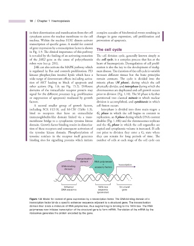
Figure 1.9 Model for control of gene expression by a transcription factor. The DNA - binding domain of a
transcription factor binds a specifi c enhancer sequence adjacent to a structural gene. The transactivation
domain then binds a molecule of RNA polymerase, thus augmenting its binding to the TATA box. The RNA
polymerase now initiates transcription of the structural gene to form mRNA. Translation of the mRNA by the
ribosomes generates the protein encoded by the gene.