
Page 26 - Essential Haematology
P. 26
12 / Chapter 1 Haemopoiesis
Death factor
APOPTOSIS e.g. Fas ligand
Caspases
Death
domain
Release of
cytochrome c
Procaspases
Increased
Inhibits BAX protein
p53
BCL-2
BAX gene
expression
Increased
BCL-2 DNA
damage
Cytotoxic drugs
Radiation
Survival factor
e.g. growth factor
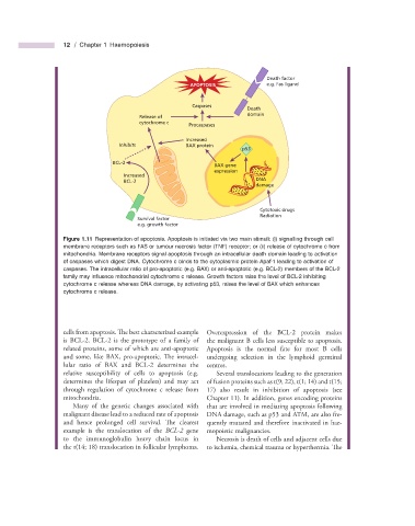
Figure 1.11 Representation of apoptosis. Apoptosis is initiated via two main stimuli: (i) signalling through cell
membrane receptors such as FAS or tumour necrosis factor (TNF) receptor; or (ii) release of cytochrome c from
mitochondria. Membrane receptors signal apoptosis through an intracellular death domain leading to activation
of caspases which digest DNA. Cytochrome c binds to the cytoplasmic protein Apaf - 1 leading to activation of
caspases. The intracellular ratio of pro - apoptotic (e.g. BAX) or anti - apoptotic (e.g. BCL - 2) members of the BCL - 2
family may infl uence mitochondrial cytochrome c release. Growth factors raise the level of BCL - 2 inhibiting
cytochrome c release whereas DNA damage, by activating p53, raises the level of BAX which enhances
cytochrome c release.
cells from apoptosis. The best characterized example Overexpression of the BCL - 2 protein makes
is BCL - 2. BCL - 2 is the prototype of a family of the malignant B cells less susceptible to apoptosis.
related proteins, some of which are anti - apoptotic Apoptosis is the normal fate for most B cells
and some, like BAX, pro - apoptotic. Th e intracel- undergoing selection in the lymphoid germinal
lular ratio of BAX and BCL - 2 determines the centres.
relative susceptibility of cells to apoptosis (e.g. Several translocations leading to the generation
determines the lifespan of platelets) and may act of fusion proteins such as t(9; 22), t(1; 14) and t(15;
through regulation of cytochrome c release from 17) also result in inhibition of apoptosis (see
mitochondria. Chapter 11 ). In addition, genes encoding proteins
Many of the genetic changes associated with that are involved in mediating apoptosis following
malignant disease lead to a reduced rate of apoptosis DNA damage, such as p53 and ATM, are also fre-
and hence prolonged cell survival. Th e clearest quently mutated and therefore inactivated in hae-
example is the translocation of the BCL - 2 gene mopoietic malignancies.
to the immunoglobulin heavy chain locus in Necrosis is death of cells and adjacent cells due
the t(14; 18) translocation in follicular lymphoma. to ischemia, chemical trauma or hyperthermia. Th e