
Page 992 - Basic _ Clinical Pharmacology ( PDFDrive )
P. 992
978 SECTION VIII Chemotherapeutic Drugs
Complement (C1–C9)
C5 → C5a, C5b B
Bacterial lysis
C5b, C6, C7, C8, C9
Chemotaxis (MAC)
C3a, C5a Eculizumab
blocks Release of salts,
cleavage of proteins, water, etc
C5
A Inflammatory 3
site
C3a, Opsonization Bacterial destruction
C5a C3b
2 Bacteria
Macrophage C3b
C3a, C 1
C5a
1
2
C3b
Monocyte Bloodstream receptor Macrophage
3
Endothelial cell
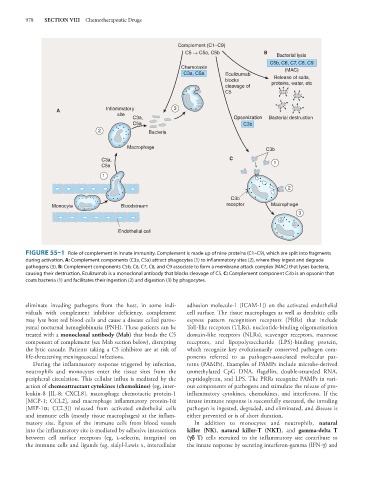
FIGURE 55–1 Role of complement in innate immunity. Complement is made up of nine proteins (C1–C9), which are split into fragments
during activation. A: Complement components (C3a, C5a) attract phagocytes (1) to inflammatory sites (2), where they ingest and degrade
pathogens (3). B: Complement components C5b, C6, C7, C8, and C9 associate to form a membrane attack complex (MAC) that lyses bacteria,
causing their destruction. Eculizumab is a monoclonal antibody that blocks cleavage of C5. C: Complement component C3b is an opsonin that
coats bacteria (1) and facilitates their ingestion (2) and digestion (3) by phagocytes.
eliminate invading pathogens from the host, in some indi- adhesion molecule-1 [ICAM-1]) on the activated endothelial
viduals with complement inhibitor deficiency, complement cell surface. The tissue macrophages as well as dendritic cells
may lyse host red blood cells and cause a disease called parox- express pattern recognition receptors (PRRs) that include
ysmal nocturnal hemoglobinuria (PNH). These patients can be Toll-like receptors (TLRs), nucleotide-binding oligomerization
treated with a monoclonal antibody (Mab) that binds the C5 domain-like receptors (NLRs), scavenger receptors, mannose
component of complement (see Mab section below), disrupting receptors, and lipopolysaccharide (LPS)-binding protein,
the lytic cascade. Patients taking a C5 inhibitor are at risk of which recognize key evolutionarily conserved pathogen com-
life-threatening meningococcal infections. ponents referred to as pathogen-associated molecular pat-
During the inflammatory response triggered by infection, terns (PAMPs). Examples of PAMPs include microbe-derived
neutrophils and monocytes enter the tissue sites from the unmethylated CpG DNA, flagellin, double-stranded RNA,
peripheral circulation. This cellular influx is mediated by the peptidoglycan, and LPS. The PRRs recognize PAMPs in vari-
action of chemoattractant cytokines (chemokines) (eg, inter- ous components of pathogens and stimulate the release of pro-
leukin-8 [IL-8; CXCL8], macrophage chemotactic protein-1 inflammatory cytokines, chemokines, and interferons. If the
[MCP-1; CCL2], and macrophage inflammatory protein-1α innate immune response is successfully executed, the invading
[MIP-1α; CCL3]) released from activated endothelial cells pathogen is ingested, degraded, and eliminated, and disease is
and immune cells (mostly tissue macrophages) at the inflam- either prevented or is of short duration.
matory site. Egress of the immune cells from blood vessels In addition to monocytes and neutrophils, natural
into the inflammatory site is mediated by adhesive interactions killer (NK), natural killer-T (NKT), and gamma-delta T
between cell surface receptors (eg, l-selectin, integrins) on (fc T) cells recruited to the inflammatory site contribute to
the immune cells and ligands (eg, sialyl-Lewis x, intercellular the innate response by secreting interferon-gamma (IFN-γ) and