
Page 993 - Basic _ Clinical Pharmacology ( PDFDrive )
P. 993
CHAPTER 55 Immunopharmacology 979
A CR ON Y MS known as CD1 and have been implicated in host defense against
microbial agents, autoimmune diseases, and tumors.
ADA Adenosine deaminase
ADC Antibody-drug conjugate The Adaptive Immune System
ALG Antilymphocyte globulin
APC Antigen-presenting cell The adaptive immune system is mobilized by cues from the innate
response when the innate processes are incapable of coping with
ATG Antithymocyte globulin
an infection. The adaptive immune system has a number of char-
CD Cluster of differentiation
acteristics that contribute to its success in eliminating pathogens.
CSF Colony-stimulating factor These include the ability to (1) respond to a variety of antigens,
CTL Cytotoxic T lymphocyte each in a specific manner; (2) discriminate between foreign (“non-
DC Dendritic cell self”) antigens (pathogens) and self antigens of the host; and
DTH Delayed-type hypersensitivity (3) respond to a previously encountered antigen in a learned way
by initiating a vigorous memory response. This adaptive response
FKBP FK-binding protein
culminates in the production of antibodies, which are the effec-
GVHD Graft-versus-host disease
tors of humoral immunity; and the activation of T lymphocytes,
HAMA Human antimouse antibody which are the effectors of cell-mediated immunity.
HLA Human leukocyte antigen The induction of specific adaptive immunity requires the par-
IFN Interferon ticipation of professional antigen-presenting cells (APCs), which
IGIV Immune globulin intravenous include dendritic cells (DCs), macrophages, and B lymphocytes.
These cells play pivotal roles in the induction of an adaptive
IL Interleukin
immune response because of their capacity to phagocytize particu-
LFA Leukocyte function-associated antigen
late antigens (eg, pathogens) or endocytose protein antigens, and
Mab Monoclonal antibody enzymatically digest them to generate peptides, which are then
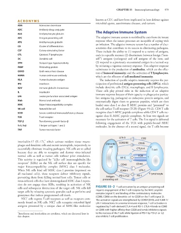
MHC Major histocompatibility complex loaded onto class I or class II MHC proteins and “presented” to
NK cell Natural killer cell the cell surface T-cell receptor (TCR) (Figure 55–2). CD8 T cells
SCID Severe combined immunodeficiency disease recognize class I–MHC peptide complexes while CD4 T cells rec-
ognize class II–MHC peptide complexes. At least two signals are
TCR T-cell receptor
necessary for the activation of T cells. The first signal is delivered
TGF-a Transforming growth factor-β
following engagement of the TCR with peptide-bound MHC
Th1, Th2 T helper cell types 1 and 2 molecules. In the absence of a second signal, the T cells become
TNF Tumor necrosis factor
LFA-3 CD2
*
interleukin-17 (IL-17), which activate resident tissue macro- CD80/86
phages and dendritic cells and recruit neutrophils, respectively, to Dendritic CD28
cell
successfully eliminate invading pathogens. NK cells are so called CTLA-4 2 T cell
because they are able to recognize and destroy virus-infected
normal cells as well as tumor cells without prior stimulation. TCR 1
This activity is regulated by “killer cell immunoglobulin-like MHC
receptors” (KIRs) on the NK cell surface that are specific for CD40 CD40L
major histocompatibility complex (MHC) class I molecules.
When NK cells bind self MHC class I proteins (expressed on ICAM-1 LFA-1
all nucleated cells), these receptors deliver inhibitory signals, T lymphocyte
preventing them from killing normal host cells. Tumor cells or Antigen- PD-L1/L2 PD-1
virus-infected cells that have downregulated MHC class I expres- presenting cell
sion do not engage these KIRs, resulting in activation of NK
cells and subsequent destruction of the target cell. NK cells kill FIGURE 55–2 T-cell activation by an antigen-presenting cell
target cells by releasing cytotoxic granules such as perforins and requires engagement of the T-cell receptor by the MHC-peptide
complex (signal 1) and binding of the costimulatory molecules
granzymes that induce programmed cell death. (CD80, CD86) on the dendritic cell to CD28 on the T cell (signal 2).
NKT cells express T-cell receptors as well as receptors com- The activation signals are strengthened by CD40/CD40L and ICAM-1/
monly found on NK cells. NKT cells recognize microbial lipid LFA-1 interactions. In a normal immune response, T-cell activation is
antigens presented by a unique class of MHC-like molecules regulated by T-cell–derived CTLA-4 and PD-1. CTLA-4 binds to CD80
or CD86 with higher affinity than CD28 and sends inhibitory signals
* Interferons and interleukins are cytokines, which are discussed later in to the nucleus of the T cell, while ligation of PD-1 by PD-L1 or -L2
this chapter. also inhibits T cell proliferation.