
Page 191 - parasitology for medical and clinical laboratoryprofessionals
P. 191
Intestinal Cestodes 171
MICROSCOPIC DIAGNOSTIC FEATURE
General Classification—Cestode “Worms”
Organism Taenia solium and Taenia saginata differentiation
Specimen Required Fecal specimen
Stage Diagnostic stages include the ova, scolex (head), or proglottids
(contains both male and female reproductive organs); the
embryonated egg is the most common stage where Taenia
species are diagnosed. The egg is yellow-brown and contains a
thick wall with radial striations.
Species differentiation is based on characteristics of proglottids,
which are used to differentiate between the Taenia sp.
(T. saginata has 15–20 uterine branches on each side of the
uterine trunk, whereas T. solium has only 7–13 uterine branches).
Size T. saginata adults reach a length of 10 m and T. solium is most
often 7 m or less.
Shape Segmented; both species are similar in shape in that they
contain a scolex and proglottids with slight differences in
internal structures.
Motility Proglottids are motile; may actively move outside of the anus.
Other Features Scolex may also be useful in addition to proglottid differences
for identifying the species; the scolex of T. saginata contains
4 suckers for attachment and that of T. solium includes a
rostellum with 25–30 hooklets along with 4 suckers.
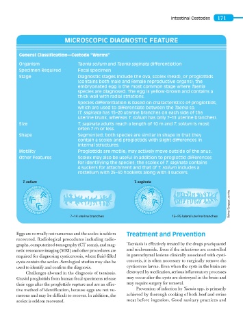
T. solium T. saginata
Delmar/Cengage Learning
7–14 uterine branches 15–25 lateral uterine branches
Eggs are normally not numerous and the scolex is seldom Treatment and Prevention
recovered. Radiological procedures including radio-
graphs, computerized tomography (CT scans), and mag- Taeniasis is effectively treated by the drugs praziquantel
netic resonance imaging (MRI) and other procedures are and niclosamide. Even if the infections are controlled
required for diagnosing cysticercosis, where fluid-filled in parenchymal lesions clinically associated with cysti-
cysts contain the scolex. Serological studies may also be cercosis, it is often necessary to surgically remove the
used to identify and confirm the diagnosis. cysticercus larvae. Even when the cysts in the brain are
Challenges abound in the diagnosis of taeniasis. destroyed by medication, serious inflammatory processes
Gravid proglottids from human fecal specimens release may occur after the cysts are destroyed in the brain and
their eggs after the proglottids rupture and are an effec- may require surgery for removal.
tive method of identification, because eggs are not nu- Prevention of infection by Taenia spp. is primarily
merous and may be difficult to recover. In addition, the achieved by thorough cooking of both beef and swine
scolex is seldom recovered. meat before ingestion. Good sanitary practices and