
Page 204 - Atlas of Histology with Functional Correlations
P. 204
damaged vessel and to release adhesive glycoproteins, adenosine
diphosphate (ADP), and serotonin. This increases the plug size by adhesion
and attraction of other platelets. The damaged endothelial cells, in turn,
release tissue factor, which initiates blood clotting, von Willebrand factor,
which facilitates adhesion of platelets to laminin and collagen in the
subendothelial tissue, and endothelin, which contracts the smooth muscle
fibers in the damaged vessel. Surface receptors on platelets bind to
fibrinogen in circulating plasma and the protein thrombin then converts
fibrinogen into solid fibrin fibrils. Fibrin forms a loose meshwork of fibrils
around the plug, trapping other platelets and blood cells to form and
strengthen the blood clot, which enlarges until bleeding stops. After blood
clot is formed and the bleeding stops, the aggregated platelets contribute to
clot retraction by pulling the damaged edges of the blood vessels together.
Following the vessel repair, the clot is removed by the proteolytic action of
the enzyme plasmin, formed from the circulating plasma protein
plasminogen.
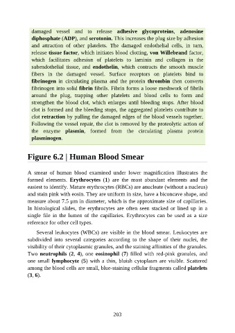
Figure 6.2 | Human Blood Smear
A smear of human blood examined under lower magnification illustrates the
formed elements. Erythrocytes (1) are the most abundant elements and the
easiest to identify. Mature erythrocytes (RBCs) are anucleate (without a nucleus)
and stain pink with eosin. They are uniform in size, have a biconcave shape, and
measure about 7.5 μm in diameter, which is the approximate size of capillaries.
In histological slides, the erythrocytes are often seen stacked or lined up in a
single file in the lumen of the capillaries. Erythrocytes can be used as a size
reference for other cell types.
Several leukocytes (WBCs) are visible in the blood smear. Leukocytes are
subdivided into several categories according to the shape of their nuclei, the
visibility of their cytoplasmic granules, and the staining affinities of the granules.
Two neutrophils (2, 4), one eosinophil (7) filled with red-pink granules, and
one small lymphocyte (5) with a thin, bluish cytoplasm are visible. Scattered
among the blood cells are small, blue-staining cellular fragments called platelets
(3, 6).
203