
Page 206 - Atlas of Histology with Functional Correlations
P. 206
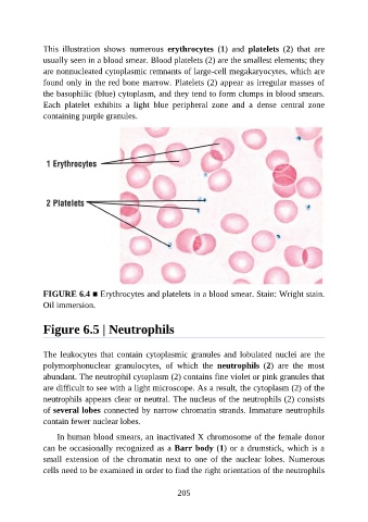
This  illustration  shows  numerous  erythrocytes  (1)  and  platelets  (2)  that  are

               usually seen in a blood smear. Blood platelets (2) are the smallest elements; they
               are nonnucleated cytoplasmic remnants of large-cell megakaryocytes, which are
               found only in the red bone marrow. Platelets (2) appear as irregular masses of
               the basophilic (blue) cytoplasm, and they tend to form clumps in blood smears.
               Each  platelet  exhibits  a  light  blue  peripheral  zone  and  a  dense  central  zone

               containing purple granules.


               FIGURE 6.4 ■ Erythrocytes and platelets in a blood smear. Stain: Wright stain.
               Oil immersion.


               Figure 6.5 | Neutrophils


               The leukocytes  that  contain  cytoplasmic  granules and  lobulated  nuclei  are  the
               polymorphonuclear  granulocytes,  of  which  the  neutrophils  (2)  are  the  most
               abundant. The neutrophil cytoplasm (2) contains fine violet or pink granules that

               are difficult to see with a light microscope. As a result, the cytoplasm (2) of the
               neutrophils appears clear or neutral. The nucleus of the neutrophils (2) consists
               of several lobes connected by narrow chromatin strands. Immature neutrophils
               contain fewer nuclear lobes.

                   In human blood smears, an inactivated X chromosome of the female donor

               can be occasionally recognized as a Barr body (1) or a drumstick, which is a
               small  extension  of  the  chromatin  next  to  one  of  the  nuclear  lobes.  Numerous
               cells need to be examined in order to find the right orientation of the neutrophils


                                                          205