
Page 205 - Atlas of Histology with Functional Correlations
P. 205
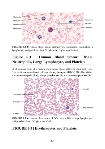
FIGURE  6.2  ■  Human  blood  smear:  erythrocytes,  neutrophils,  eosinophils,  a

               lymphocyte, and platelets. Stain: Wright stain. High magnification.


               Figure  6.3  |  Human  Blood  Smear:  RBCs,


               Neutrophils, Large Lymphocyte, and Platelets


               A photomicrograph of a human blood smear shows different blood cell types.
               The most numerous blood cells are the erythrocytes (RBCs) (1). Also visible
               are two neutrophils (2, 4), a large lymphocyte (5), and numerous platelets (3).


               FIGURE  6.3  ■  Human  blood  smear:  RBCs,  neutrophils,  a  large  lymphocyte,
               and platelets. Stain: Wright stain. ×205.


               FIGURE 6.4 | Erythrocytes and Platelets


                                                          204