
Page 210 - Atlas of Histology with Functional Correlations
P. 210
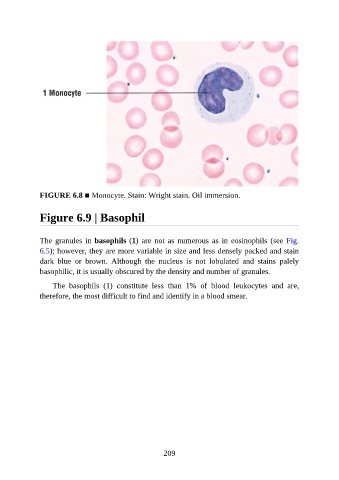
FIGURE 6.8 ■ Monocyte. Stain: Wright stain. Oil immersion.


               Figure 6.9 | Basophil


               The granules in basophils (1) are not as numerous as in eosinophils (see Fig.
               6.5); however, they are more variable in size and less densely packed and stain

               dark  blue  or  brown.  Although  the  nucleus  is  not  lobulated  and  stains  palely
               basophilic, it is usually obscured by the density and number of granules.

                   The  basophils  (1)  constitute  less  than  1%  of  blood  leukocytes  and  are,
               therefore, the most difficult to find and identify in a blood smear.


                                                          209