Page 208 - Atlas of Histology with Functional Correlations
P. 208
macrophages in the spleen, liver, and bone marrow.


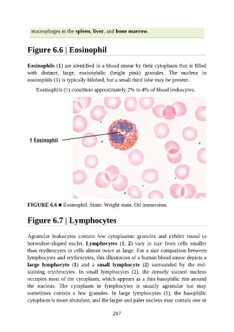
               Figure 6.6 | Eosinophil


               Eosinophils (1) are identified in a blood smear by their cytoplasm that is filled
               with  distinct,  large,  eosinophilic  (bright  pink)  granules.  The  nucleus  in

               eosinophils (1) is typically bilobed, but a small third lobe may be present.

                   Eosinophils (1) constitute approximately 2% to 4% of blood leukocytes.


               FIGURE 6.6 ■ Eosinophil. Stain: Wright stain. Oil immersion.


               Figure 6.7 | Lymphocytes


               Agranular  leukocytes  contain  few  cytoplasmic  granules  and  exhibit  round  to
               horseshoe-shaped  nuclei.  Lymphocytes  (1,  2)  vary  in  size  from  cells  smaller

               than erythrocytes to cells almost twice as large. For a size comparison between
               lymphocytes and erythrocytes, this illustration of a human blood smear depicts a
               large  lymphocyte  (1)  and  a  small  lymphocyte  (2)  surrounded  by  the  red-

               staining  erythrocytes.  In  small  lymphocytes  (2),  the  densely  stained  nucleus
               occupies most of the cytoplasm, which appears as a thin basophilic rim around
               the  nucleus.  The  cytoplasm  in  lymphocytes  is  usually  agranular  but  may
               sometimes  contain  a  few  granules.  In  large  lymphocytes  (1),  the  basophilic
               cytoplasm is more abundant, and the larger and paler nucleus may contain one or


                                                          207

